 Back
BackThe Integumentary System: Structure and Function
Study Guide - Smart Notes
 Tailored notes based on your materials, expanded with key definitions, examples, and context.
The Integumentary System
Overview
The integumentary system is the body's largest organ system, serving as a protective barrier and playing key roles in sensation, thermoregulation, and metabolic functions. It is composed of the cutaneous membrane (skin) and accessory structures such as nails, hair, and exocrine glands.
Cutaneous membrane: Includes the epidermis and dermis.
Accessory structures: Comprise nails, hair, and exocrine glands (sebaceous and sudoriferous glands).
Cell Types of the Epidermis
Main Cell Types
Keratinocytes: Produce keratin, a fibrous protein that strengthens and waterproofs the skin. These cells form the majority of the epidermis and are also found in hair and nails.
Melanocytes: Located at the base of the epidermis, these cells synthesize melanin, the pigment responsible for skin color and protection against ultraviolet (UV) radiation.
Langerhans cells: Specialized immune cells (macrophages) that originate from bone marrow and participate in immune defense by interacting with T helper cells.
Merkel cells: Sensory receptors associated with nerve endings, responsible for detecting touch and pressure.
Layers of the Epidermis
Stratification of the Epidermis
The epidermis is organized into distinct layers, each with specialized functions. From deep to superficial, the layers are:
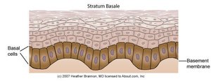
Stratum basale (germinativum): The deepest layer, consisting of a single row of stem cells that continuously divide to produce new keratinocytes. This layer is essential for epidermal regeneration.
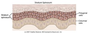
Stratum spinosum: Several layers of polygonal keratinocytes that begin synthesizing keratin. Cells in this layer are connected by desmosomes, giving a spiny appearance.
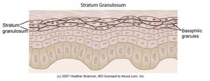
Stratum granulosum: Cells lose their nuclei and accumulate keratohyalin granules and lipids, contributing to the skin's waterproof barrier.
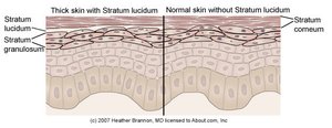
Stratum lucidum: A thin, clear layer found only in thick skin (e.g., palms, soles). It provides an additional barrier to reduce friction.
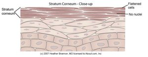
Stratum corneum: The outermost layer, composed of dead, flattened keratinocytes (corneocytes) filled with keratin. This layer protects against abrasion and dehydration.
Mnemonic: "Come, Let's Get Sun Burned" (Corneum, Lucidum, Granulosum, Spinosum, Basale)
Stratum Basale
The stratum basale is the site of active cell division, providing new cells to replenish the epidermis. It contains melanocytes and Merkel cells.
Stratum Spinosum
Cells in the stratum spinosum transition from columnar to polygonal shape and begin keratin synthesis. Langerhans cells are also present here.
Stratum Granulosum
This layer is characterized by the presence of basophilic granules and the production of keratin and lipids, which are essential for the skin's barrier function.
Stratum Lucidum
The stratum lucidum is only present in thick skin and serves to reduce friction between the stratum corneum and stratum granulosum.
Stratum Corneum
The stratum corneum consists of dead, flattened cells that provide a durable overcoat for the body, preventing water loss and entry of pathogens.
Skin Color
Pigments Contributing to Skin Color
Melanin: Produced by melanocytes; color ranges from yellow to black. Increased melanin production occurs with sun exposure, providing some protection against UV damage.
Carotene: A yellow-orange pigment found in the stratum corneum and fat tissue.
Hemoglobin: The red pigment in blood, visible in fair skin due to low melanin content.
Excessive sun exposure can damage skin, leading to elastin clumping, immune suppression, and increased cancer risk.
The Dermis
Structure and Function
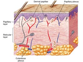
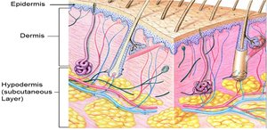
The dermis is a strong, flexible connective tissue layer beneath the epidermis. It contains fibroblasts, macrophages, mast cells, and white blood cells, as well as collagen, elastin, and reticular fibers. The dermis houses blood vessels, nerves, hair follicles, and glands.
Papillary layer: The upper layer, composed of loose connective tissue and thin collagen fibers. Contains dermal papillae that increase surface area for exchange of gases, nutrients, and waste products.
Reticular layer: The deeper, thicker layer made of dense irregular connective tissue with thick collagen fibers, providing strength and elasticity.
Accessory Structures
Exocrine Glands
Sebaceous (Oil) Glands: Secrete sebum into hair follicles or directly onto the skin. Sebum lubricates and waterproofs skin and hair, and inhibits bacterial growth. Absent on palms and soles.
Sudoriferous (Sweat) Glands: Two main types:
Apocrine glands: Found in armpits, pubic region, and areolae; secrete a viscous fluid into hair follicles.
Merocrine (Eccrine) glands: Distributed throughout most of the skin; secrete watery sweat directly onto the skin surface, important for thermoregulation and excretion.
Nails
Nails are scale-like modifications of the epidermis that protect the distal phalanges. The nail matrix at the base is responsible for nail growth, and the nail body slides over the nail bed as it grows. Nails are heavily keratinized and changes in their appearance can indicate systemic health issues.
Subcutaneous Layer (Hypodermis)
Structure and Function
The subcutaneous layer, or hypodermis, is composed of loose connective tissue and adipose tissue. It anchors the skin to underlying structures, acts as a shock absorber, and insulates the body. Approximately half of the body's fat is stored here. The hypodermis is not considered part of the skin proper.
Summary Table: Layers of the Epidermis
Layer | Location | Main Features |
|---|---|---|
Stratum corneum | Outermost | Dead, keratinized cells; barrier function |
Stratum lucidum | Thick skin only | Clear, thin layer; reduces friction |
Stratum granulosum | Middle | Keratohyalin granules; lipid production |
Stratum spinosum | Above basale | Keratin synthesis; spiny appearance |
Stratum basale | Deepest | Stem cells; melanocytes; cell division |
Additional info: Hair follicles and hair are distributed over most of the body and provide minor protective functions, but are not covered in detail here as per the study guide instructions.
