 Back
BackThe Integumentary System: Structure, Function, and Clinical Relevance
Study Guide - Smart Notes
 Tailored notes based on your materials, expanded with key definitions, examples, and context.
The Integumentary System
Overview and Terminology
The integumentary system is the body's largest organ system, primarily composed of the skin and its associated structures. It serves as a protective barrier, regulates temperature, and facilitates sensation and communication. Understanding key terms is essential for grasping the clinical and physiological aspects of this system.
Apoptosis: Programmed cell death, a natural process for removing unneeded or abnormal cells. Blocked in cancer cells.
Dermatitis: Inflammation of the skin.
Scab: Dried blood clot that forms over a wound, protecting underlying tissue during healing.
Scar tissue: Formed with fewer collagen fibers and blood vessels at the repair site.
Regenerative: Ability to restore or renew tissue after damage.
Necrosis: Death of cells or tissue due to injury, infection, or lack of blood flow.
Metastasis: Spread of cancer cells from the original site to other body parts.
Benign: Non-cancerous mass of abnormal cells; does not spread.
Malignant: Cancerous, invasive, and capable of spreading.
Oncogenic: Gene mutations that cause cancer.
Debridement: Removal of necrotic tissue to promote healing.
Lesion: Area of tissue injury.
Erythema: Redness of the skin due to increased blood flow.
Hematoma: Localized collection of blood outside blood vessels, causing discoloration.
Cyanosis: Bluish skin color due to low oxygen levels.
Pallor: Paleness, often due to anemia or decreased blood flow.
Wrinkles and sagging: Result from thinning epidermis and decreased elastin in elderly individuals.
Organs and Structures of the Integumentary System
Skin (Cutaneous Membrane)
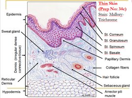
The skin is the largest organ in the body and consists of two main layers: the epidermis and dermis, separated by a basement membrane. It is classified as a cutaneous membrane, one of the four main body membranes.
Epidermis: Outermost layer, composed of stratified squamous keratinized epithelial tissue. It is avascular and consists of 4-5 layers depending on skin thickness.
Dermis: Deeper layer, made of connective tissue, vascularized, and provides nutrients to the epidermis. It contains two sublayers: papillary (areolar connective tissue) and reticular (dense irregular connective tissue with collagen and elastin fibers).
Hypodermis (Subcutaneous Layer): Not a true skin layer but associated with it. Composed mainly of adipose tissue, providing insulation and energy storage.
Associated Structures
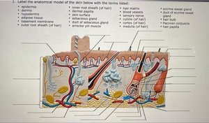
Hair (Pilus): Three types: lanugo (infant), vellus (child, thin), terminal (coarse, adult). Alopecia refers to hair loss; male pattern baldness is a sex-linked trait.
Nails: Composed of the nail edge, matrix, cuticle, and lunula.
Glands:
Apocrine: Sweat glands in armpits, groin; active at puberty.
Merocrine (Eccrine): Most numerous, especially on palms and soles; secrete watery sweat.
Sebaceous: Oil glands associated with hair follicles.
Ceruminous: Produce earwax (cerumen).
Layers of the Skin
Epidermal Layers (Superficial to Deep)
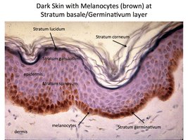
Stratum Corneum: Outermost, dead keratinized cells; site of callus and corn formation.
Stratum Lucidum: Present only in thick skin (palms, soles).
Stratum Granulosum: Keratinocytes accumulate keratin and melanin.
Stratum Spinosum: Provides strength and flexibility.
Stratum Basale (Germinativum): Deepest, mitotically active; contains melanocytes.
Thick skin is found on palms and soles; thin skin is found on eyelids and lips.
Cells of the Epidermis
Melanocytes: Produce melanin pigment, found in the stratum basale.
Keratinocytes: Most abundant; produce keratin for protection.
Tactile (Merkel) Cells: Sensory receptors for touch.
Langerhans (Dendritic) Cells: Immune defense cells.
Dermal Layers
Papillary Dermis: Areolar connective tissue, contains capillaries and sensory neurons.
Reticular Dermis: Dense irregular connective tissue, provides strength and elasticity.
Stretch marks result from torn collagen fibers; tension lines follow the orientation of collagen bundles.
Functions of the Skin
Vitamin D Production: UV light stimulates synthesis of cholecalciferol, converted to calcitriol by the liver and kidneys, essential for calcium and phosphate absorption.
Communication: Expresses emotions and health status.
Sensation: Contains receptors for touch, pain, temperature.
Nutrient Storage: Stores lipids and minerals.
Excretion: Removes salts and wastes via sweat.
Protection: Shields underlying tissues from mechanical, chemical, and biological harm.
Temperature Regulation: Sweat glands and arrector pili muscles (cause goosebumps) help maintain body temperature.
Immunity: Epidermal cells and macrophages provide innate immune defense.
Skin Pigmentation and Clinical Color Changes
Melanin: Brown-black pigment produced by melanocytes; amount determines skin color.
Carotene: Yellow-orange pigment from diet (e.g., carrots).
Hemoglobin: Red pigment in blood, contributes to skin color.
All humans have similar numbers of melanocytes, but melanin production varies. Blood flow and oxygenation also affect skin color:
Erythema: Redness from increased blood flow.
Cyanosis: Blue tint from low oxygen.
Pallor: Paleness from reduced blood flow or anemia.
Jaundice: Yellowing from bilirubin accumulation.
Vitiligo: Loss of melanocytes, causing patchy skin color.
Albinism: Absence of melanin production.
Wound Healing and Burns
Wound Healing Steps
Hemostasis: Stop bleeding (clot formation).
Inflammation: Immune response to clear debris and pathogens.
Proliferation: Growth and rebuilding of tissue.
Remodeling: Strengthening and maturation of new tissue.
Burns
First-degree: Involves only the epidermis.
Second-degree: Involves epidermis and part of dermis (partial thickness).
Third-degree: Extends through dermis to underlying tissues (full thickness); may require grafting.
Types of grafts include autograft (self), allograft (another human), and xenograft (another species). The greatest immediate threat in severe burns is dehydration.
Skin Cancer
Basal Cell Carcinoma: Most common, least dangerous.
Melanoma: Most dangerous, arises from melanocytes.
Summary Table: Layers and Structures of the Skin
Layer | Main Tissue | Key Structures |
|---|---|---|
Epidermis | Stratified squamous epithelium | Keratinocytes, melanocytes, Merkel cells, Langerhans cells |
Dermis (Papillary) | Areolar connective tissue | Capillaries, sensory neurons |
Dermis (Reticular) | Dense irregular connective tissue | Collagen, elastin, hair follicles, glands |
Hypodermis | Adipose tissue | Fat storage, insulation |
Additional info:
Vitamin D synthesis begins in the skin and is completed in the liver and kidneys, forming calcitriol, which regulates calcium homeostasis.
Clinical assessment of burns often uses the "rule of nines" to estimate the affected body surface area.
