 Back
BackThe Muscular System: Structure and Function
Study Guide - Smart Notes
 Tailored notes based on your materials, expanded with key definitions, examples, and context.
The Muscular System
Overview of Muscle Anatomy
The muscular system is composed of skeletal muscles, which are responsible for movement, posture, and heat production. Muscles are attached to bones and work together with the skeletal system to facilitate voluntary movements. The arrangement and structure of muscles vary throughout the body, reflecting their specific functions.
Skeletal muscles are striated, voluntary muscles attached to the skeleton.
Muscle fibers are the basic units of muscle tissue, organized into bundles called fascicles.
Tendons connect muscles to bones, transmitting the force generated by muscle contraction.
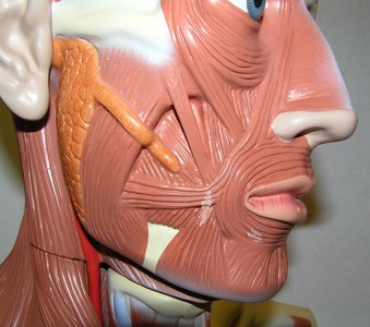
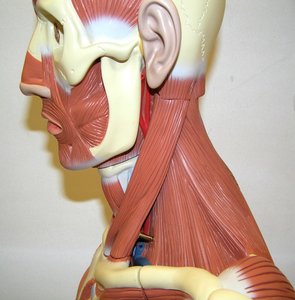
Muscles of the Head and Neck
The head and neck contain numerous muscles responsible for facial expression, mastication (chewing), and movement of the head. These muscles are intricately arranged and often overlap.
Facial muscles allow for expressions such as smiling, frowning, and blinking.
Masticatory muscles include the masseter and temporalis, which are essential for chewing.
Neck muscles such as the sternocleidomastoid facilitate head rotation and flexion.
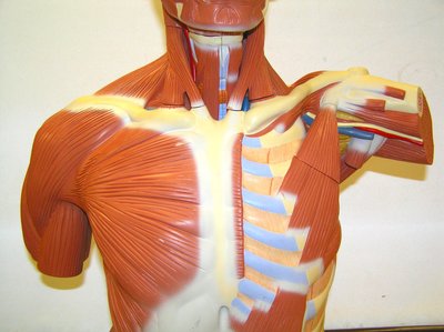
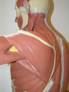
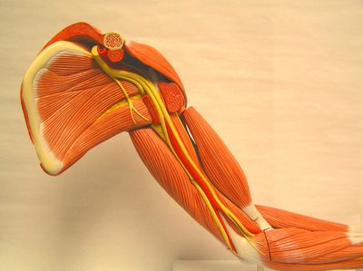
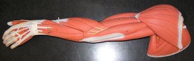
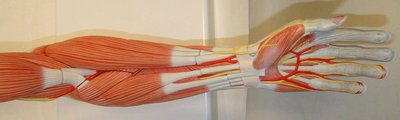
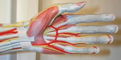
Muscles of the Upper Limb
The upper limb muscles are specialized for a wide range of movements, including lifting, pushing, and fine motor control. These muscles are grouped into those acting on the shoulder, arm, forearm, and hand.
Shoulder muscles include the deltoid, rotator cuff group, and pectoralis major, which stabilize and move the shoulder joint.
Arm muscles such as the biceps brachii and triceps brachii are responsible for flexion and extension of the elbow.
Forearm and hand muscles control wrist and finger movements, enabling gripping and manipulation.
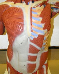
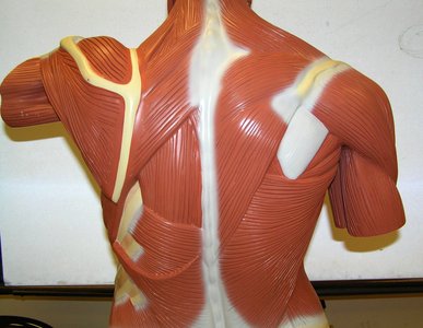
Muscles of the Back
The back muscles are essential for maintaining posture and enabling movements such as extension, rotation, and lateral flexion of the spine. The erector spinae group is the primary muscle group responsible for these actions.
Erector spinae muscles include the iliocostalis, longissimus, and spinalis, which extend and stabilize the vertebral column.
Quadratus lumborum assists in lateral flexion of the spine.
Multifidus provides stability to the vertebral column.
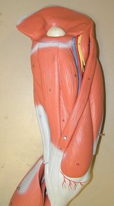
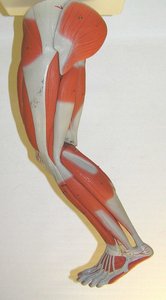
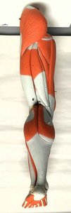
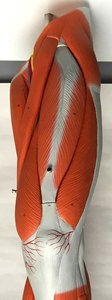
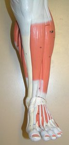
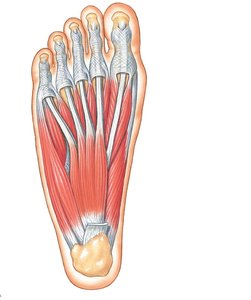
Muscles of the Lower Limb
The lower limb muscles are adapted for locomotion, weight-bearing, and balance. They are organized into groups acting on the hip, thigh, leg, and foot.
Hip muscles such as the gluteus maximus, medius, and minimus are responsible for extension, abduction, and rotation of the thigh.
Thigh muscles include the quadriceps (anterior) and hamstrings (posterior), which extend and flex the knee, respectively.
Leg muscles such as the gastrocnemius and tibialis anterior control movements of the ankle and foot.
Foot muscles enable toe movements and maintain the arches of the foot.

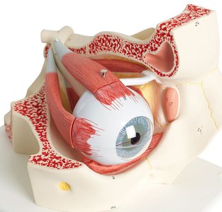
Muscles of the Eye
The eye is moved by six extrinsic muscles, which allow for precise control of eye position and movement. These muscles are anchored to the orbital bones and insert onto the sclera of the eyeball.
Rectus muscles (superior, inferior, medial, lateral) move the eye in straight directions.
Oblique muscles (superior and inferior) rotate the eye.
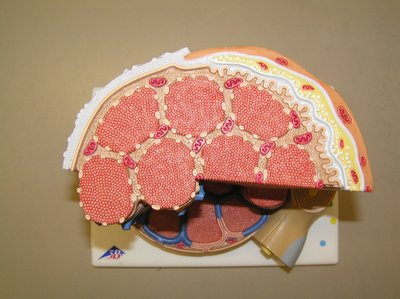
Microscopic Structure of Skeletal Muscle
Skeletal muscle tissue is composed of long, cylindrical fibers organized into fascicles. Each fiber contains myofibrils, which are made up of repeating units called sarcomeres. The sarcomere is the functional unit of muscle contraction.
Myofibrils contain actin and myosin filaments responsible for contraction.
Sarcomere is the basic contractile unit, defined by Z-lines.
Muscle contraction occurs via the sliding filament mechanism.
Major Muscle Groups and Their Functions
Muscle groups are classified based on their location and function. Understanding these groups is essential for identifying their roles in movement and stability.
Flexors decrease the angle at a joint (e.g., biceps brachii).
Extensors increase the angle at a joint (e.g., triceps brachii).
Abductors move a limb away from the midline (e.g., deltoid).
Adductors move a limb toward the midline (e.g., adductor longus).
Summary Table: Major Muscle Groups and Actions
Muscle Group | Location | Primary Action |
|---|---|---|
Deltoid | Shoulder | Abduction of arm |
Biceps brachii | Upper arm | Flexion of elbow |
Triceps brachii | Upper arm | Extension of elbow |
Quadriceps femoris | Thigh | Extension of knee |
Hamstrings | Thigh | Flexion of knee |
Gastrocnemius | Calf | Plantar flexion of foot |
Gluteus maximus | Hip | Extension of thigh |
Rectus abdominis | Abdomen | Flexion of vertebral column |
Muscle Contraction: The Sliding Filament Theory
Muscle contraction is explained by the sliding filament theory, which describes how actin and myosin filaments interact to shorten the sarcomere and produce force.
ATP provides energy for myosin heads to bind and pull actin filaments.
Calcium ions released from the sarcoplasmic reticulum initiate contraction.
Relaxation occurs when calcium is reabsorbed and ATP binds to myosin, breaking the cross-bridge.
Equation:
Clinical Relevance
Understanding muscle anatomy is essential for diagnosing and treating musculoskeletal disorders, injuries, and conditions such as muscular dystrophy, strains, and sprains.
Muscle injuries often involve tears or strains of muscle fibers or tendons.
Rehabilitation focuses on restoring muscle strength and function.
Conclusion
The muscular system is integral to movement, stability, and overall function of the human body. Mastery of muscle anatomy and physiology is foundational for students in anatomy and physiology courses.
