 Back
BackThe Nervous System: Structure, Function, and Protection
Study Guide - Smart Notes
 Tailored notes based on your materials, expanded with key definitions, examples, and context.
The Nervous System: Overview
Main Divisions of the Nervous System
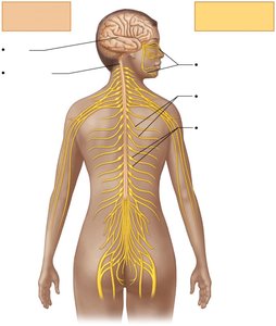
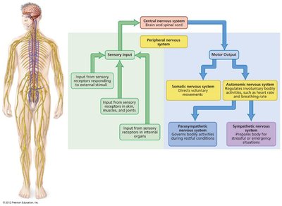
The nervous system is the master control and communication system of the body. It is divided into two main anatomical divisions: the Central Nervous System (CNS) and the Peripheral Nervous System (PNS).
Central Nervous System (CNS): Consists of the brain and spinal cord. It is the integration and command center, interpreting sensory input and dictating motor output.
Peripheral Nervous System (PNS): Consists of cranial nerves, spinal nerves, and ganglia. It carries messages to and from the CNS, serving as communication lines that link all parts of the body to the CNS.
Functional Organization
The nervous system has three overlapping functions:
Sensory Input: Gathering information from sensory receptors about internal and external changes.
Integration: Processing and interpreting sensory input to decide what should be done at each moment.
Motor Output: Activating effector organs (muscles and glands) to cause a response.
Nervous Tissue
Types of Tissue in the Nervous System
Nervous tissue is specialized for communication and consists of two main cell types: neurons and neuroglia (supporting cells).
Neurons: Excitable cells that transmit electrical signals.
Neuroglia: Non-excitable cells that support, protect, and insulate neurons.
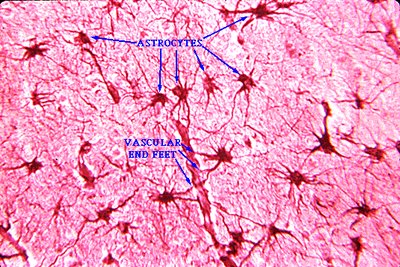
Neuroglia (Glial Cells)
Types and Functions of Neuroglia in the CNS
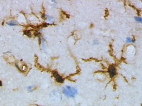
Neuroglia in the CNS include astrocytes, microglial cells, ependymal cells, and oligodendrocytes. Each type has specialized functions:
Astrocytes: Most abundant; support neurons, regulate the blood-brain barrier, and participate in information processing.
Microglial Cells: Monitor neuron health, act as phagocytes to remove debris and pathogens.
Ependymal Cells: Line brain ventricles and spinal cord; circulate cerebrospinal fluid (CSF).
Oligodendrocytes: Produce myelin sheaths in the CNS, insulating axons and speeding impulse transmission.

Neuroglia in the PNS
Satellite Cells: Surround neuron cell bodies in the PNS; function similarly to astrocytes.
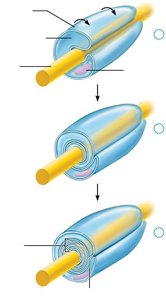
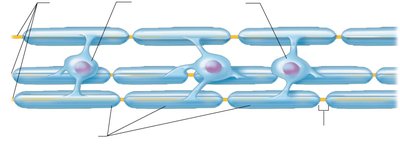
Schwann Cells: Form myelin sheaths around peripheral nerve fibers; vital for regeneration of damaged nerves.

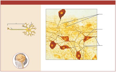
Neurons: Structure and Function
General Characteristics
Neurons are the structural and functional units of the nervous system. They are highly specialized for conducting impulses and have the following properties:
Extreme longevity: Can function for a lifetime.
Amitotic: Most do not divide after development (few exceptions).
High metabolic rate: Require continuous supply of oxygen and glucose.
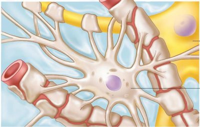
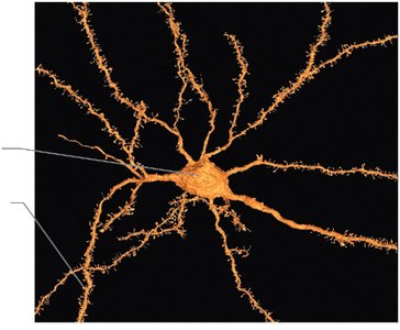
Neuron Structure
Cell Body (Soma): Contains the nucleus and organelles; biosynthetic center of the neuron.
Dendrites: Short, branching processes; receptive/input regions that convey signals toward the cell body.
Axon: Single long process; generates and transmits nerve impulses away from the cell body. Axons may branch (axon collaterals) and end in axon terminals.
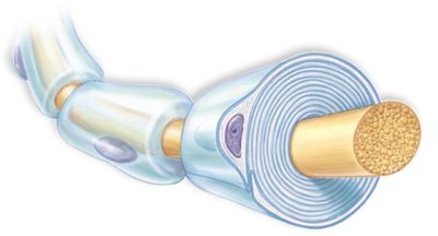
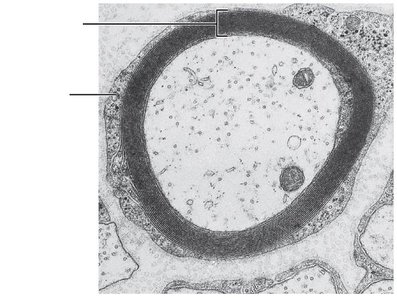
Myelin Sheath
The myelin sheath is a segmented, whitish, protein-lipid covering that insulates axons and increases the speed of nerve impulse transmission.
Myelinated fibers: Conduct impulses rapidly.
Nonmyelinated fibers: Conduct impulses more slowly.
Nodes of Ranvier: Gaps in the myelin sheath where axon membrane is exposed; facilitate rapid signal conduction.
Functional Classification of Neurons
Types of Neurons
Sensory (Afferent) Neurons: Transmit impulses from sensory receptors toward the CNS; mostly unipolar, cell bodies in ganglia of PNS.
Motor (Efferent) Neurons: Carry impulses from the CNS to effectors (muscles/glands); multipolar, cell bodies in CNS.
Interneurons (Association Neurons): Lie between sensory and motor neurons; most abundant, multipolar, and found within CNS.
Major Regions of the Brain
Brain Regions and Functions
The adult brain is divided into four main regions:
Cerebrum: Largest part; responsible for higher brain functions such as thought, memory, and voluntary movement.
Diencephalon: Includes thalamus, hypothalamus, and epithalamus; involved in sensory relay, homeostasis, and endocrine function.
Cerebellum: Coordinates movement and balance.
Brain Stem: Includes midbrain, pons, and medulla oblongata; controls automatic behaviors necessary for survival.
Cerebral Cortex
The cerebral cortex is the outer layer of the cerebrum, responsible for conscious thought, sensory perception, voluntary motor initiation, communication, memory, and understanding.
Gyri: Ridges on the brain surface.
Sulci: Shallow grooves.
Fissures: Deep grooves separating major brain regions.
Lobes of the Cerebrum
Frontal Lobe: Voluntary movement, planning, reasoning, problem-solving.
Parietal Lobe: Sensory perception and integration.
Temporal Lobe: Hearing, memory, language.
Occipital Lobe: Vision.
Insula: Taste, visceral sensation, and autonomic control.
Functional Areas of the Cortex
Motor Areas: Control voluntary movement (e.g., primary motor cortex, premotor cortex, Broca's area).
Sensory Areas: Receive and interpret sensory information (e.g., primary somatosensory cortex, visual cortex).
Association Areas: Integrate information for purposeful action (e.g., prefrontal cortex, Wernicke's area).
Protection of the Brain
Meninges
The brain is protected by three connective tissue membranes called meninges:
Dura Mater: Strongest, outermost layer; has periosteal and meningeal layers.
Arachnoid Mater: Middle layer with web-like extensions; subarachnoid space contains CSF and blood vessels.
Pia Mater: Delicate, innermost layer; clings tightly to the brain surface and contains many blood vessels.
Dural septa partition the cranial cavity and limit brain movement. Dural venous sinuses collect venous blood from the brain and drain into the jugular veins.
Cerebrospinal Fluid (CSF)
CSF is a clear, colorless fluid that surrounds the brain and spinal cord, providing buoyancy, protection, and chemical stability.
Formation: Produced by the choroid plexus in the ventricles of the brain.
Circulation: Flows through ventricles, subarachnoid space, and central canal of the spinal cord; reabsorbed into venous blood via arachnoid granulations.
Functions: Cushions the brain, reduces its effective weight, nourishes neural tissue, and removes waste products.
Summary Table: Main Neuroglia of the CNS and PNS
Neuroglia Type | Location | Main Function |
|---|---|---|
Astrocytes | CNS | Support neurons, regulate blood-brain barrier, maintain environment |
Microglial Cells | CNS | Phagocytosis of debris and pathogens |
Ependymal Cells | CNS | Line ventricles, circulate CSF |
Oligodendrocytes | CNS | Form myelin sheaths around CNS axons |
Satellite Cells | PNS | Support neuron cell bodies in ganglia |
Schwann Cells | PNS | Form myelin sheaths around PNS axons, aid regeneration |
Key Equations and Concepts
Resting Membrane Potential: (Nernst equation for potassium)
Ohm's Law (for neurons): , where is current, is voltage, and is resistance.
Clinical Application
Multiple Sclerosis (MS): An autoimmune disease where the immune system attacks myelin sheaths in the CNS, leading to impaired nerve conduction, muscle weakness, and sensory disturbances.
Example: A patient with MS may experience visual disturbances, muscle weakness, and loss of coordination due to demyelination of CNS axons.
Additional info: This guide covers the structure and function of the nervous system, neuroglia, neuron anatomy, brain regions, and protective mechanisms, as well as clinical relevance for exam preparation.
