 Back
BackThe Respiratory System: Structure and Function
Study Guide - Smart Notes
 Tailored notes based on your materials, expanded with key definitions, examples, and context.
The Respiratory System
Overview
The respiratory system is essential for gas exchange, supplying oxygen to the body and removing carbon dioxide. It consists of a series of organs and structures that facilitate the movement and exchange of gases between the atmosphere and the bloodstream.
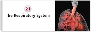
Anatomy of the Respiratory System
Main Components
Nose and Nasal Cavity: Entryway for air, encased in cranial and facial bones.
Pharynx (Throat): Passageway for air, food, and liquids.
Larynx (Voice Box): Located in the anterior neck, responsible for sound production and protecting the airway.
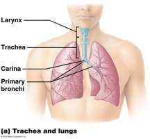
Trachea (Windpipe): Conducts air to the lungs, located in the mediastinum.
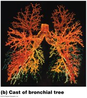
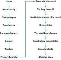
Bronchial Tree: Branching tubes that distribute air to the lungs.
Lungs: Spongy organs containing millions of alveoli for gas exchange.
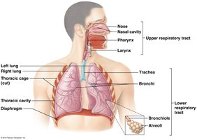
Respiratory Tract Divisions
Upper Respiratory Tract: Extends from the nasal cavity to the larynx.
Lower Respiratory Tract: Extends from the trachea to the alveoli.
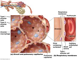
Alveoli: Tiny air sacs where gas exchange occurs.
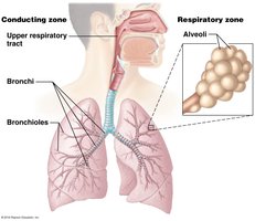
Functional Zones of the Respiratory System
Conducting Zone
The conducting zone consists of all the respiratory passages that carry air to the sites of gas exchange. It includes the nose, nasal cavity, pharynx, larynx, trachea, bronchi, and bronchioles. Its main functions are to filter, warm, and moisten incoming air.
Respiratory Zone
The respiratory zone is where actual gas exchange occurs. It includes structures that contain alveoli, such as respiratory bronchioles, alveolar ducts, and alveolar sacs.
The Nose and Nasal Cavity
External and Internal Structures
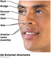
External Structures: Include the root, bridge, dorsum nasi, apex, alae, and anterior nares (nostrils).
Internal Structures: Composed of nasal bones, lateral cartilages, septal cartilage, alar cartilages, and dense connective tissue.
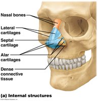
Nasal Cavity Anatomy
Nasal Conchae: Superior, middle, and inferior conchae increase surface area and help warm and humidify air.
Nasal Meatuses: Passageways beneath each concha.
Olfactory Mucosa: Contains receptors for the sense of smell.
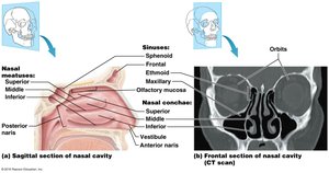
The Pharynx
Divisions of the Pharynx
Nasopharynx: Posterior to the nasal cavity; passageway for air only.
Oropharynx: Posterior to the oral cavity; passageway for air, food, and liquids.
Laryngopharynx: Extends from the hyoid bone to the esophagus; passageway for air and food.
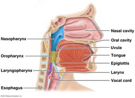
The Larynx
Cartilages of the Larynx
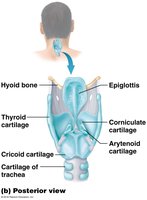
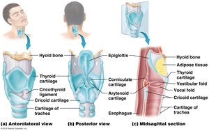
Thyroid Cartilage: Largest cartilage, forms the anterior and superior walls; known as the Adam's apple.
Cricoid Cartilage: Inferior to the thyroid cartilage; provides support and attachment for ligaments and muscles.
Epiglottis: Elastic cartilage that covers the glottis during swallowing to prevent food from entering the airway.
Arytenoid, Corniculate, and Cuneiform Cartilages: Paired cartilages involved in sound production and support.
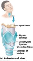
Vocal Folds and Sound Production
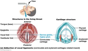
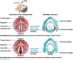
Vestibular Folds (False Vocal Cords): Close off the glottis during swallowing; do not produce sound.
True Vocal Cords: Vibrate to produce sound as air passes over them; pitch and loudness are controlled by tension and force of air.
The Trachea and Bronchial Tree
Trachea
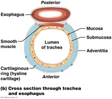
Structure: Supported by C-shaped rings of hyaline cartilage, which keep the airway open.
Carina: Last tracheal cartilage ring; contains sensory receptors that trigger the cough reflex.
Bronchial Tree
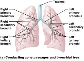
Primary Bronchi: Right and left branches entering each lung.
Secondary (Lobar) Bronchi: Three on the right, two on the left, each serving a lung lobe.
Tertiary (Segmental) Bronchi: Further divisions supplying bronchopulmonary segments.
Bronchioles: Smallest airways leading to alveolar ducts and alveoli.
Alveoli and the Respiratory Membrane
Cell Types in Alveoli
Type I Alveolar Cells: Squamous cells for rapid gas diffusion.
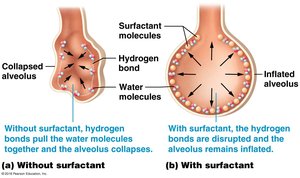
Type II Alveolar Cells: Produce surfactant to reduce surface tension and prevent alveolar collapse.
Alveolar Macrophages: Phagocytes that remove debris and pathogens.
The Lungs and Pleurae
Lung Structure
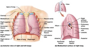
Lobes: Right lung has three lobes; left lung has two lobes and a cardiac notch for the heart.
Hilum: Entry and exit point for bronchi, blood vessels, lymphatics, and nerves.
Pleurae: Double-layered serous membranes (visceral and parietal) that reduce friction and create a pressure gradient for lung inflation.
Pulmonary Ventilation
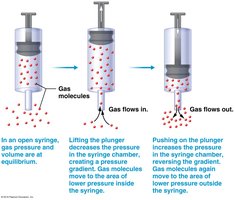
Pressure-Volume Relationship (Boyle's Law)
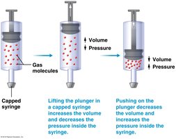
Boyle’s law states that at a constant temperature, the pressure and volume of a gas are inversely related:
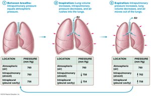
As lung volume increases, pressure decreases, allowing air to flow in (inspiration).
As lung volume decreases, pressure increases, pushing air out (expiration).
Mechanics of Breathing
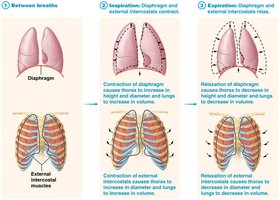
Inspiration: Diaphragm and external intercostal muscles contract, increasing thoracic volume and decreasing pressure.
Expiration: Usually passive; diaphragm and external intercostals relax, decreasing thoracic volume and increasing pressure.
Physical Factors Influencing Ventilation
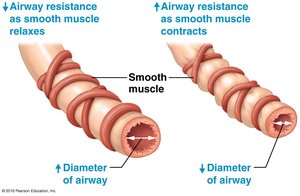
Airway Resistance: Determined by airway diameter; bronchodilation decreases resistance, bronchoconstriction increases resistance.
Alveolar Surface Tension: Surfactant reduces surface tension, preventing alveolar collapse (atelectasis).
Pulmonary Compliance: The ability of the lungs and chest wall to stretch.
Pulmonary Volumes and Capacities
Key Volumes
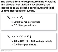
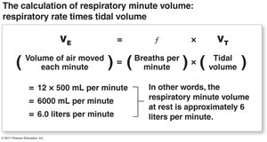
Tidal Volume (TV): Air inspired or expired during normal breathing (~500 mL).
Inspiratory Reserve Volume (IRV): Additional air that can be inhaled after a normal inspiration.
Expiratory Reserve Volume (ERV): Additional air that can be exhaled after a normal expiration.
Residual Volume (RV): Air remaining in lungs after maximal expiration.
Gas Exchange
Dalton’s Law of Partial Pressures
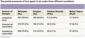
Each gas in a mixture exerts its own pressure (partial pressure). The total pressure is the sum of all partial pressures.
Henry’s Law
The amount of gas dissolved in a liquid is proportional to its partial pressure and solubility in the liquid.
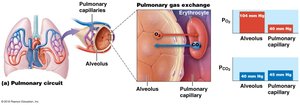
Pulmonary Gas Exchange (External Respiration)
Oxygen diffuses from alveoli (high PO2) to blood (low PO2).
Carbon dioxide diffuses from blood (high PCO2) to alveoli (low PCO2).
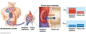
Tissue Gas Exchange (Internal Respiration)
Oxygen diffuses from blood to tissues (where PO2 is lower).
Carbon dioxide diffuses from tissues (where PCO2 is higher) to blood.
Oxygen Transport
Hemoglobin and Oxygen Saturation
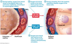
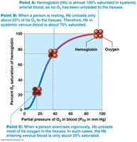
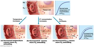
Most oxygen is transported bound to hemoglobin (Hb) in erythrocytes.
Each Hb molecule can bind up to four oxygen molecules.
Percent saturation depends on PO2 and Hb affinity for oxygen.
Carbon Dioxide Transport
Transport Mechanisms
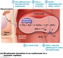
7–10% dissolved in plasma
20% bound to hemoglobin (carbaminohemoglobin)
70% as bicarbonate ions (HCO3–) in plasma, formed by the reaction:
Neural Control of Ventilation
Respiratory Centers
Medulla Oblongata: Contains the respiratory rhythm generator (RRG), ventral respiratory group (VRG), and dorsal respiratory group (DRG).
Pons: Modifies respiratory rhythm.
Chemoreceptor Regulation
Central Chemoreceptors: Located in the medulla; respond to changes in CO2 and H+ in cerebrospinal fluid.
Peripheral Chemoreceptors: Located in carotid and aortic bodies; respond to changes in blood PO2, PCO2, and pH.
Summary Table: Partial Pressures of Gases in Air
Source of Sample | Nitrogen (N2) | Oxygen (O2) | Carbon Dioxide (CO2) | Water Vapor (H2O) |
|---|---|---|---|---|
Inhaled air (dry) | 597 (78.6%) | 159 (20.9%) | 0.3 (0.04%) | 3.7 (0.5%) |
Alveolar air (saturated) | 573 (75.4%) | 100 (13.2%) | 40 (5.2%) | 47 (6.2%) |
Exhaled air (saturated) | 569 (74.8%) | 116 (15.3%) | 28 (3.7%) | 47 (6.2%) |
Additional info: This table summarizes the partial pressures of the main gases in different air samples, illustrating the changes that occur during respiration.
