 Back
BackThe Respiratory System: Structure, Function, and Physiology
Study Guide - Smart Notes
 Tailored notes based on your materials, expanded with key definitions, examples, and context.
The Respiratory System
Overview and Functions
The respiratory system is essential for gas exchange, sound production, and olfaction. It provides a large surface area for the exchange of oxygen and carbon dioxide between the air and blood, protects respiratory surfaces, and helps regulate airflow and defense mechanisms.
Gas Exchange: Facilitates the movement of O2 into the blood and CO2 out of the blood.
Air Movement: Moves air to and from the lungs for efficient gas exchange.
Protection: Defends against dehydration, temperature changes, and pathogens.
Sound Production: Vocal cords in the larynx produce sounds for speech.
Olfaction: Olfactory receptors in the nasal cavity detect odors.

Organization of the Respiratory System
Upper and Lower Respiratory Tracts
The respiratory system is divided into upper and lower tracts, each with distinct anatomical structures and functions.
Upper Respiratory System: Nose, nasal cavity, paranasal sinuses, and pharynx. Responsible for filtering, warming, and humidifying incoming air.
Lower Respiratory System: Larynx, trachea, bronchi, bronchioles, and alveoli. Conducts air to the lungs and is the site of gas exchange.
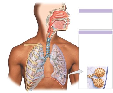
Respiratory Tract: Conducting and Respiratory Portions
The respiratory tract is functionally divided into conducting and respiratory portions:
Conducting Portion: Extends from the nasal cavity to the larger bronchioles; moves air to the sites of gas exchange.
Respiratory Portion: Includes the smallest bronchioles and alveoli; where gas exchange occurs.
Alveoli: Tiny air sacs in the lungs where all gas exchange takes place.
Respiratory Mucosa and Defense System
Structure of Respiratory Mucosa
The respiratory mucosa lines the conducting portion of the respiratory tract and consists of an epithelium and an underlying layer of areolar tissue (lamina propria). In the upper tract, mucous glands secrete mucus onto the epithelial surface, while in the lower tract, smooth muscle cells encircle the bronchioles.
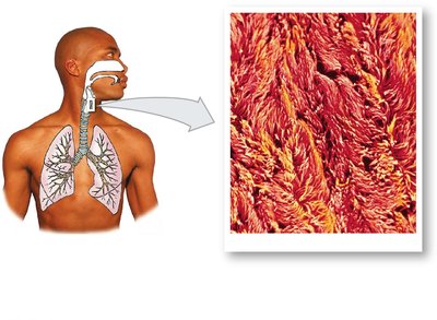
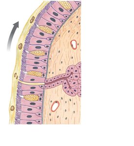
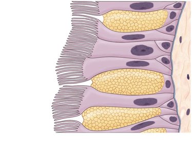
Respiratory Defense Mechanisms
The respiratory defense system protects the lungs from pathogens and debris:
Filtration: Nasal hairs trap large particles in the nasal vestibule.
Mucus: Produced by mucous cells and glands, traps smaller particles and microorganisms.
Cilia: Move mucus and trapped debris toward the pharynx to be swallowed.
Alveolar Macrophages: Engulf small particles that reach the alveoli.
Upper Respiratory System
Nose and Nasal Cavity
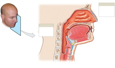
The nose is the primary entryway for air. Air passes through the nostrils (nares) into the nasal vestibule, where nasal hairs filter large particles. The nasal septum divides the cavity, and the superior region contains olfactory receptors for smell. Mucus from paranasal sinuses cleans and moistens the air.
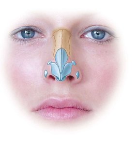
Air Flow and Meatuses
Air flows from the vestibule through the superior, middle, and inferior nasal meatuses, which create turbulence to trap particles, warm and humidify air, and direct olfactory stimuli to receptors.
Palates and Pharynx
Hard Palate: Forms the floor of the nasal cavity, separating it from the oral cavity.
Soft Palate: Extends behind the hard palate, dividing the nasopharynx from the rest of the pharynx.
Pharynx: Shared by the respiratory and digestive systems, divided into nasopharynx, oropharynx, and laryngopharynx.
Lower Respiratory System
Larynx
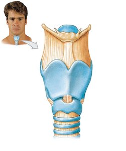
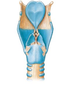
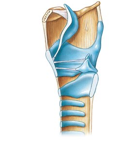
The larynx is a cartilaginous structure that surrounds and protects the glottis (the opening between the vocal cords). Major cartilages include the thyroid, cricoid, and epiglottis. The epiglottis prevents food and liquids from entering the airway during swallowing. Smaller cartilages (arytenoid, corniculate, cuneiform) help open/close the glottis and produce sound.
Sound Production
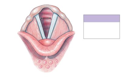
Sound is produced as air passes through the glottis, vibrating the vocal folds to create sound waves.
Trachea and Bronchial Tree
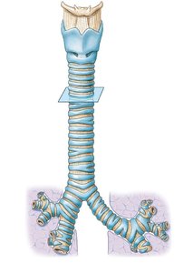
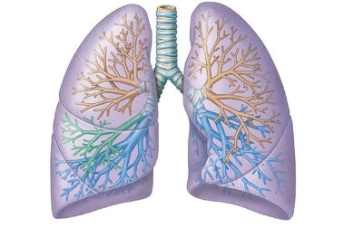
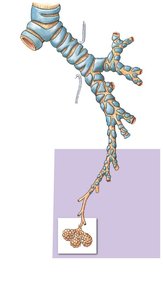
The trachea (windpipe) is a tough, flexible tube that branches into the right and left main bronchi. Each main bronchus divides into lobar bronchi (supplying lung lobes), which further branch into segmental bronchi and then bronchioles. Bronchioles lack cartilage and are dominated by smooth muscle, which regulates airflow resistance.
Alveoli and Gas Exchange Structures
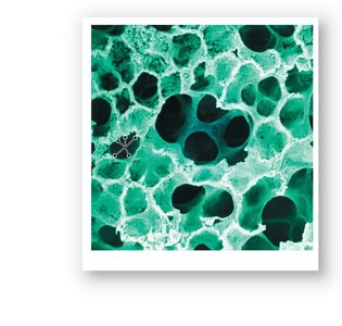
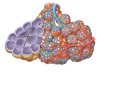
Terminal bronchioles branch into respiratory bronchioles, which connect to alveolar ducts ending in alveolar sacs. Each alveolus is surrounded by capillaries and elastic fibers, forming the primary site of gas exchange.
Surfactant and Blood Air Barrier
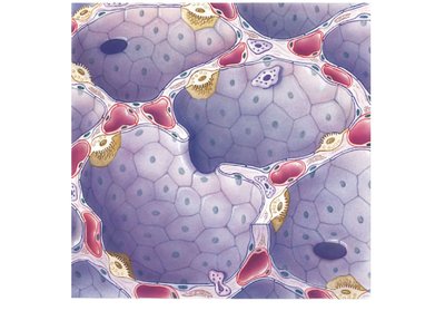
Surfactant, an oily secretion produced by alveolar cells, reduces surface tension and prevents alveolar collapse. Gas exchange occurs across the blood air barrier, which consists of the alveolar cell layer, capillary endothelium, and a fused basement membrane.
The Lungs
Gross Anatomy
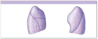
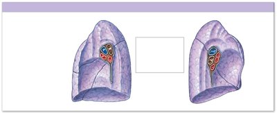
The right lung has three lobes (superior, middle, inferior) separated by horizontal and oblique fissures. The left lung has two lobes (superior, inferior) separated by an oblique fissure and features a cardiac notch. Lungs are divided into smaller compartments by trabeculae and interlobular septa.
Pleura and Pleural Cavities
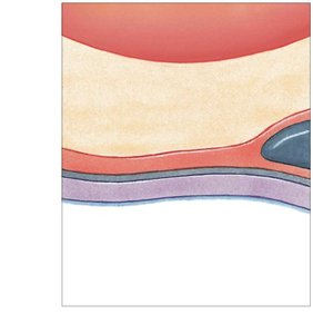
Pleura: Serous membrane with two layers—parietal (lining thoracic wall) and visceral (covering lungs).
Pleural Fluid: Lubricates the space between pleural layers, reducing friction during breathing.
Respiration: External and Internal
Definitions and Processes
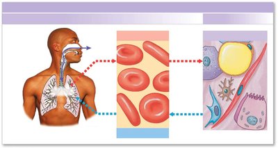
External Respiration: Exchange of O2 and CO2 between interstitial fluid and the external environment, primarily via pulmonary ventilation (breathing).
Internal Respiration: Exchange of O2 and CO2 between interstitial fluid and cells, involving cellular respiration.
Pulmonary Ventilation
Mechanics of Breathing
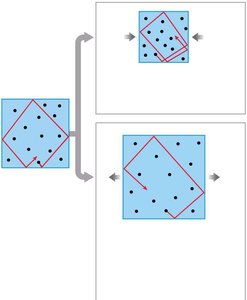
Pulmonary ventilation is driven by pressure gradients created by changes in thoracic volume. Air moves from areas of higher to lower pressure. The relationship between pressure and volume is described by Boyle's Law:
Boyle's Law:
Decreasing volume increases pressure; increasing volume decreases pressure.
Inhalation and Exhalation
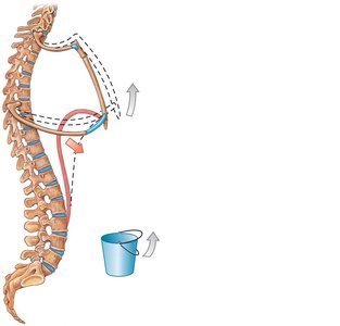
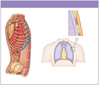
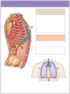
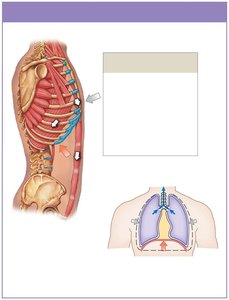
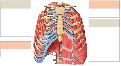
During inhalation, the diaphragm and external intercostal muscles contract, increasing thoracic volume and decreasing pressure, causing air to flow in. During exhalation, these muscles relax, thoracic volume decreases, pressure increases, and air flows out. Inhalation is always active; exhalation can be passive or active (using accessory muscles).
Intrapleural Pressure and Respiratory Pump
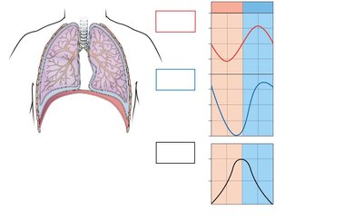
Intrapleural pressure (between pleural layers) remains below atmospheric pressure, creating a respiratory pump that assists venous return to the heart. Cyclical changes in this pressure are essential for normal breathing.
Tidal Volume and Dead Space
Tidal Volume: The amount of air moved into or out of the lungs during a single respiratory cycle (about 500 mL in adults).
Anatomic Dead Space: Portion of inhaled air that remains in conducting passages and does not participate in gas exchange.
Gas Exchange and Transport
Principles of Gas Exchange
Gas exchange occurs across the blood air barrier and depends on partial pressures and solubility of gases. Diffusion occurs in response to concentration gradients, and the efficiency of exchange is enhanced by short diffusion distances, large surface area, and coordinated blood/air flow.
Oxygen and Carbon Dioxide Transport
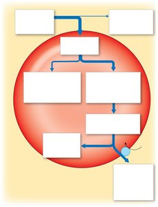
Oxygen: Most O2 is transported bound to hemoglobin in red blood cells as oxyhemoglobin (HbO2).
Carbon Dioxide: Transported in three forms: dissolved in plasma, bound to hemoglobin (carbaminohemoglobin), or converted to carbonic acid (H2CO3), which dissociates into H+ and HCO3-.
Control and Integration of Respiration
Regulation of Breathing
Peripheral and Alveolar Capillaries: Adjust oxygen delivery and ventilation-perfusion ratio to maintain efficient gas exchange.
Neural Control: Respiratory centers in the brainstem regulate the rate and depth of breathing. Emotional states and anticipation of exercise can alter respiratory patterns via the autonomic nervous system.
Integration with Cardiovascular System
The respiratory and cardiovascular systems work together to maintain homeostasis of O2 and CO2 in peripheral tissues. Chemoreceptors and baroreceptors coordinate changes in respiratory rate, blood pressure, and cardiac output to optimize gas exchange.
Additional info: This guide expands on the provided lecture slides with definitions, examples, and academic context to ensure a comprehensive understanding of the respiratory system for Anatomy & Physiology students.
