 Back
BackThe Skeletal System: Structure and Function (Chapter 7 Study Notes)
Study Guide - Smart Notes
 Tailored notes based on your materials, expanded with key definitions, examples, and context.
The Skeletal System
Overview of the Skeleton
The human skeleton is a complex framework of approximately 206 bones and associated cartilages. It provides support, protection, and facilitates movement. The skeleton is divided into two main divisions: the axial skeleton and the appendicular skeleton.
Axial Skeleton: Forms the longitudinal axis of the body and is structured primarily for protection. It includes the skull, vertebral column, and thoracic cage.
Appendicular Skeleton: Structured for motion and includes the bones of the girdles and the upper and lower limbs.
Main regions of the skeleton:
Skull: 22 bones (8 cranial, 14 facial)
Vertebral Column: 33 vertebrae (cervical, thoracic, lumbar, sacrum, coccyx)
Thoracic Cage: 12 pairs of ribs, sternum, part of vertebral column
Pectoral Girdle: Clavicle and scapula
Upper Limb: Humerus, radius, ulna, carpals, metacarpals, phalanges
Pelvic Girdle: Two pelvic bones (ilium, ischium, pubis) and sacrum
Lower Limb: Femur, tibia, fibula, tarsals, metatarsals, phalanges
Bone Markings
Bones display various surface features called bone markings that serve as sites for muscle, ligament, and tendon attachment, or as passages for nerves and blood vessels.
Depressions: Allow passage of blood vessels and nerves or articulation between bones.
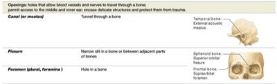
Openings: Permit access for delicate structures to pass through bones.
Projections: Sites for ligament and tendon attachment or articulation with other bones.
Type | Description | Example |
|---|---|---|
Canal (meatus) | Tunnel through a bone | Temporal bone: external acoustic meatus |
Fissure | Narrow slit in a bone or between adjacent parts of bones | Sphenoid bone: superior orbital fissure |
Foramen | Hole in a bone | Frontal bone: supraorbital foramen |
Skull Structure
Overview of Skull Bones
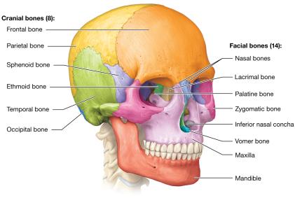
The skull is the most complex structure in the skeleton, consisting of cranial and facial bones. In adults, all skull bones are united by immovable joints called sutures, except for the mandible.
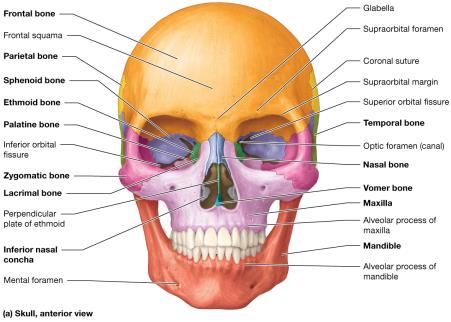
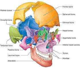
Cranial Bones (8): Frontal, parietal (2), temporal (2), occipital, sphenoid, ethmoid
Facial Bones (14): Maxilla (2), zygomatic (2), nasal (2), lacrimal (2), palatine (2), inferior nasal concha (2), vomer, mandible
Major Cavities of the Skull
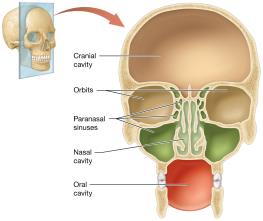
The skull contains several important cavities that house and protect sensory organs and the brain:
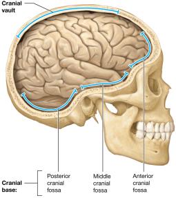
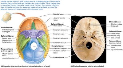
Cranial Cavity: Surrounds the brain, divided into the cranial vault (calvaria) and cranial base. The base contains the anterior, middle, and posterior cranial fossae, which support the brain.
Orbits: House the eyeballs and associated structures.
Nasal Cavity: Contains sensory receptors for smell and forms the first part of the respiratory tract.
Oral Cavity: Houses the teeth and tongue, forming the first part of the digestive tract.
Paranasal Sinuses: Air-filled spaces that lighten the skull and enhance voice resonance.
Detailed Structure of Cranial and Facial Bones
Each cranial and facial bone has unique features and articulations. The following tables summarize the main bones and their characteristics:
Bone | Description |
|---|---|
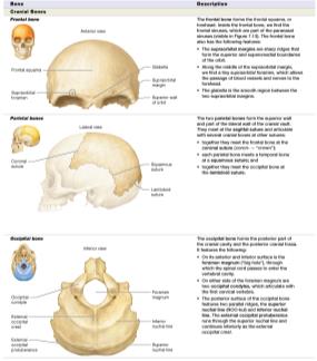
Frontal | Forms the forehead and superior part of the orbit |
Parietal | Form the superior and lateral walls of the cranium |
Occipital | Forms the posterior part and base of the skull |
Bone | Description |
|---|---|
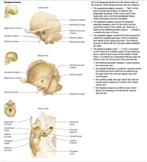
Temporal | Form the lateral walls of the cranium; house structures of the ear |
Bone | Description |
|---|---|
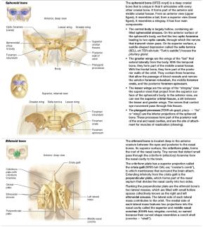
Sphenoid | Bat-shaped bone; forms part of the cranial floor |
Ethmoid | Located deep in the anterior cranium; forms part of the nasal septum and orbit |
Bone | Description |
|---|---|
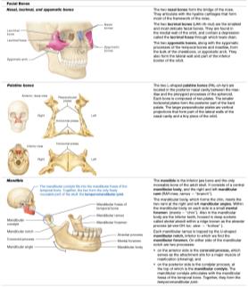
Nasal | Form the bridge of the nose |
Lacrimal | Form part of the medial wall of the orbit |
Zygomatic | Form the cheekbones |
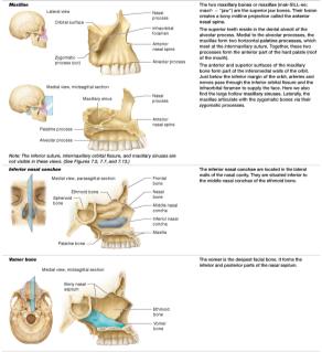
Palatine | Form part of the hard palate |
Mandible | Lower jaw bone; only movable bone of the skull |
Bone | Description |
|---|---|
Maxilla | Form the upper jaw and part of the hard palate |
Vomer | Forms the inferior part of the nasal septum |
Views of the Skull
Understanding the skull requires examining it from multiple perspectives:
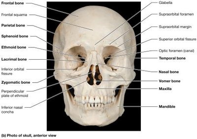
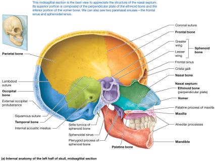
Anterior View: Shows the frontal, nasal, maxilla, zygomatic, and mandible bones.
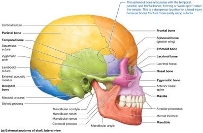
Lateral View: Highlights the temporal, sphenoid, and parietal bones, as well as the zygomatic arch.
Posterior and Superior Views: Reveal the occipital and parietal bones and the major sutures.
Inferior View: Shows the base of the skull, including the foramen magnum and occipital condyles.
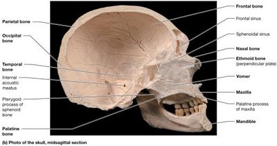
Midsagittal Section: Displays the internal structure, including the nasal septum and paranasal sinuses.
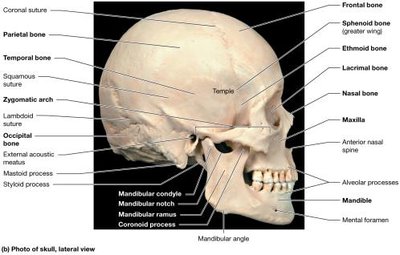
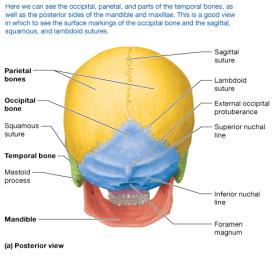
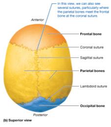
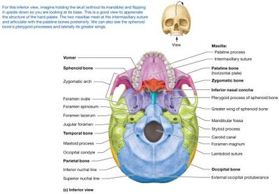
Cavities of the Skull
The Orbit
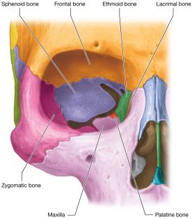
The orbit houses the eyeball and associated structures. It is formed by seven bones: frontal, maxilla, zygomatic, sphenoid, ethmoid, lacrimal, and palatine.
The Nasal Cavity
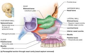
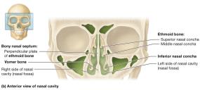
The nasal cavity is the first part of the respiratory tract and is formed by several bones lined with mucous membranes. The nasal septum divides the cavity into two fossae. Deviations in the septum can affect breathing.
Paranasal Sinuses
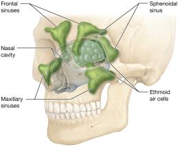
Paranasal sinuses are air-filled spaces within the frontal, sphenoid, ethmoid, and maxillary bones. They lighten the skull and enhance voice resonance. Infections can cause sinusitis.
Oral Cavity
The oral cavity houses the teeth, tongue, and some salivary glands. It is formed by the hard palate (roof), maxillae and mandible (walls), and soft tissues (floor and posterior wall).
Fetal Skull
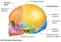
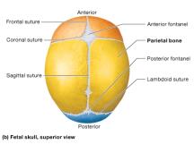
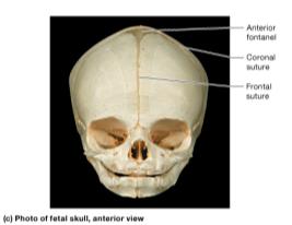
The fetal skull contains membranous areas called fontanels where ossification is incomplete. These "soft spots" provide flexibility during birth and close by 18–24 months of age.
Anterior Fontanel: Between frontal and parietal bones
Posterior Fontanel: Between parietal and occipital bones
Sphenoid Fontanels: At the temples
Mastoid Fontanels: At the junction of parietal, temporal, and occipital bones
Forensic Skull Anatomy
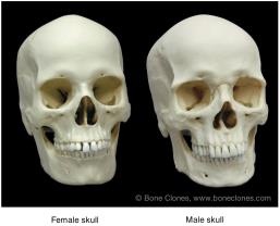
Skull bones can be used to determine a person's sex, age, and sometimes ethnic heritage. Key differences include the slope of the forehead, prominence of the supraorbital ridge, mandibular angle, and size of the mastoid process.
Hyoid Bone
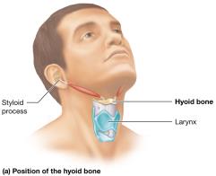
The hyoid bone is a small, C-shaped bone suspended in the superior neck by muscles and ligaments. It serves as an attachment point for muscles involved in swallowing and speech and does not articulate with any other bone.
The Vertebral Column
Overview and Structure
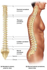
The vertebral column, or spine, consists of 33 vertebrae divided into cervical, thoracic, lumbar, sacral, and coccygeal regions. It supports the head, protects the spinal cord, and provides attachment points for ribs and muscles.
Cervical: 7 vertebrae (C1–C7)
Thoracic: 12 vertebrae (T1–T12)
Lumbar: 5 vertebrae (L1–L5)
Sacrum: 5 fused vertebrae
Coccyx: 3–5 fused vertebrae
Spinal curvatures include primary (thoracic and sacral, present at birth) and secondary (cervical and lumbar, develop after birth).
Abnormal Spinal Curvatures
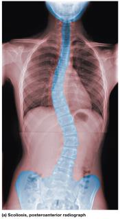
Scoliosis: Lateral curvature (C or S shaped); may be congenital, neuromuscular, or idiopathic.
Lordosis: Exaggerated cervical and lumbar curvatures; also called "swayback."
Kyphosis: Exaggerated thoracic curvature; also called "humpback."
Structure of a Typical Vertebra
All vertebrae share common features:
Body (Centrum): Weight-bearing region
Vertebral Foramen: Passage for the spinal cord
Pedicles and Laminae: Form the vertebral arch
Articular Processes: Form joints with adjacent vertebrae
Transverse and Spinous Processes: Sites for muscle attachment
Specialized Vertebrae
Cervical Vertebrae: Smallest; C1 (atlas) supports the skull, C2 (axis) allows rotation.
Thoracic Vertebrae: Articulate with ribs; heart-shaped bodies.
Lumbar Vertebrae: Largest and strongest; kidney-shaped bodies.
Sacrum and Coccyx: Fused vertebrae forming the posterior pelvic wall and tailbone.
Intervertebral Discs and Herniated Discs
Intervertebral discs are pads of fibrocartilage between vertebrae, consisting of a jelly-like nucleus pulposus and a tough anulus fibrosus. Herniated discs occur when the nucleus pulposus protrudes through a tear in the anulus fibrosus, causing nerve compression and pain.
The Thoracic Cage
Structure and Function
The thoracic cage consists of the sternum, 12 pairs of ribs, and thoracic vertebrae. It protects the heart, lungs, and major blood vessels.
Sternum: Manubrium, body, xiphoid process
Ribs: 1–7 are true ribs, 8–12 are false ribs (11–12 are floating ribs)
The Pectoral Girdle and Upper Limb
Pectoral Girdle
Composed of the clavicle and scapula, the pectoral girdle supports the upper limb and allows for a wide range of motion.
Upper Limb Bones
Humerus: Only bone of the arm; articulates with the scapula at the shoulder and with the radius and ulna at the elbow.
Radius and Ulna: Bones of the forearm; articulate with each other and with the humerus and carpals.
Carpals: Eight short bones of the wrist.
Metacarpals and Phalanges: Bones of the hand and fingers.
The Pelvic Girdle and Lower Limb
Pelvic Girdle
The pelvic girdle consists of two pelvic bones and the sacrum. Each pelvic bone is formed by the fusion of the ilium, ischium, and pubis. The pelvis supports the weight of the upper body and protects pelvic organs.
Lower Limb Bones
Femur: Thigh bone; largest and strongest bone in the body.
Patella: Kneecap; a sesamoid bone within the quadriceps tendon.
Tibia and Fibula: Bones of the leg; tibia is medial and weight-bearing, fibula is lateral.
Tarsals: Seven short bones of the ankle.
Metatarsals and Phalanges: Bones of the foot and toes.
Study Boost: Mnemonics and Visual Analogies
PEST OF 6: Parietal, Ethmoid, Sphenoid, Temporal, Occipital, Frontal (cranial bones)
Breakfast at 7, Lunch at 12, Dinner at 5: 7 cervical, 12 thoracic, 5 lumbar vertebrae
Some Lovers Try Positions That They Can't Handle: Scaphoid, Lunate, Triquetrum, Pisiform, Trapezium, Trapezoid, Capitate, Hamate (carpal bones)
