Back
BackThe Skeletal System: Structure, Function, and Development
Study Guide - Smart Notes
 Tailored notes based on your materials, expanded with key definitions, examples, and context.
The Skeletal System
Overview and Components
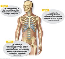
The skeletal system forms the internal framework of the body, providing structure, protection, and facilitating movement. It is composed of bones, joints, cartilages, and ligaments.
Bones (skeleton): The rigid organs that form the majority of the skeletal system.
Joints: Articulations where two or more bones meet, allowing for movement.
Cartilages: Flexible connective tissues found in joints, ear, nose, and other structures.
Ligaments: Strong bands of connective tissue that connect bones to other bones.
Subdivisions of the Skeleton
Axial skeleton: Forms the longitudinal axis of the body (skull, vertebral column, bony thorax).
Appendicular skeleton: Composed of the limbs and girdles (pectoral and pelvic girdles).
Functions of Bones
Support: Provides a framework that supports the body and cradles soft organs.
Protection: Protects vital organs (e.g., skull protects the brain, rib cage protects thoracic organs).
Movement: Skeletal muscles attach to bones, using them as levers to produce movement.
Mineral and Fat Storage: Stores minerals such as calcium and phosphorus, and fats in the internal marrow cavity.
Blood Cell Formation (Hematopoiesis): Occurs within the marrow cavities of certain bones.
Classification of Bones
Types of Osseous Tissue
Compact bone: Dense, smooth, and homogeneous tissue.
Spongy bone: Composed of small needlelike pieces (trabeculae) and many open spaces.
Bone Shapes
Long bones: Longer than they are wide; mostly compact bone (e.g., femur, humerus).
Short bones: Generally cube-shaped; mostly spongy bone (e.g., carpals, tarsals).
Flat bones: Thin, flattened, and usually curved; two thin layers of compact bone sandwiching spongy bone (e.g., skull, ribs, sternum).
Irregular bones: Do not fit into other categories (e.g., vertebrae, hip bones).
Structure of Bone
Long Bone Anatomy
Diaphysis: Shaft of the bone, composed of compact bone.
Periosteum: Fibrous connective tissue membrane covering the diaphysis, secured by Sharpey's fibers.
Epiphysis: Ends of the bone, composed mostly of spongy bone enclosed by a thin layer of compact bone.
Articular cartilage: Covers the external surface of the epiphyses; made of hyaline cartilage to decrease friction at joints.
Epiphyseal plate/line: Plate of hyaline cartilage in growing bone (for lengthwise growth); becomes the epiphyseal line in adults.
Endosteum: Lines the inner surface of the shaft; made of connective tissue.
Medullary cavity: Central cavity; contains yellow marrow (fat) in adults and red marrow (for blood cell formation) in children.
Bone Markings
Projections/processes: Sites of muscle, tendon, and ligament attachment; all begin with "T".
Depressions/cavities: Indentations; all begin with "F" (except facet).
Microscopic Anatomy
Osteon (Haversian system): Structural and functional unit of compact bone, consisting of a central canal surrounded by concentric lamellae.
Osteocytes: Mature bone cells located in lacunae.
Lamellae: Concentric rings of bone matrix.
Canaliculi: Tiny canals connecting lacunae to each other and to the central canal.
Perforating (Volkmann's) canals: Run perpendicular to the central canals, connecting blood and nerve supply.
Bone Formation, Growth, and Remodeling
Ossification
Ossification is the process of bone formation, occurring on hyaline cartilage models or fibrous membranes. Two major phases in long bones:
Osteoblasts cover hyaline cartilage with bone matrix.
Cartilage is digested away, opening up a medullary cavity.
Growth and Remodeling
New cartilage forms on the external face of articular cartilages and epiphyseal plates; old cartilage is replaced by bone matrix.
Appositional growth increases bone width.
Bone growth is regulated by hormones (growth hormone, sex hormones).
Bones are remodeled throughout life in response to blood calcium levels and mechanical stress.
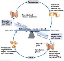
Calcium Homeostasis
Parathyroid hormone (PTH): Released when blood calcium is low; activates osteoclasts to release calcium from bone.
Calcitonin: Released when blood calcium is high; stimulates calcium deposition in bone.
Axial Skeleton
Major Components
Skull: Formed by cranial and facial bones; protects the brain and forms the structure of the face.
Vertebral column: Provides axial support; consists of 26 vertebrae separated by intervertebral discs.
Bony thorax (thoracic cage): Protects organs of the thoracic cavity; includes sternum, ribs, and thoracic vertebrae.
Skull
8 cranial bones and 14 facial bones.
Bones joined by sutures (immovable joints), except the mandible.
Paranasal sinuses lighten the skull and amplify sounds.
Hyoid bone does not articulate with any other bone; aids in swallowing and speech.
Vertebral Column
7 cervical, 12 thoracic, 5 lumbar vertebrae; sacrum (5 fused), coccyx (3–5 fused).
Primary curvatures (thoracic, sacral) present from birth; secondary curvatures (cervical, lumbar) develop after birth.
Thoracic Cage
Consists of sternum, ribs (true, false, floating), and thoracic vertebrae.
Appendicular Skeleton
Major Components
Pectoral girdle: Clavicle and scapula; attaches upper limbs to axial skeleton.
Pelvic girdle: Two coxal bones, sacrum, and coccyx; supports the weight of the upper body and protects pelvic organs.
Limbs: Upper (humerus, radius, ulna, carpals, metacarpals, phalanges) and lower (femur, tibia, fibula, tarsals, metatarsals, phalanges).
Pelvic Girdle Differences
Female pelvis is wider, shallower, and lighter for childbirth.
Joints (Articulations)
Functions and Classifications
Hold bones together and allow for mobility.
Functional classification: Synarthroses (immovable), amphiarthroses (slightly movable), diarthroses (freely movable).
Structural classification: Fibrous (immovable), cartilaginous (immovable/slightly movable), synovial (freely movable).
Synovial Joints
Articulating bones separated by a joint cavity filled with synovial fluid.
Features: articular cartilage, articular capsule, joint cavity, reinforcing ligaments.
Types: plane, hinge, pivot, condylar, saddle, ball-and-socket joints.
Developmental Aspects of the Skeleton
From Birth to Adulthood
Fetal long bones are initially hyaline cartilage; flat bones are fibrous membranes.
As the fetus grows, bone models are converted to bone.
At birth, the head and trunk are proportionately longer than the lower limbs.
During puberty, the female pelvis broadens and the male skeleton becomes more robust.
By the end of adolescence, epiphyseal plates are fully ossified.
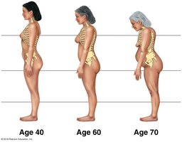
Aging and the Skeleton
Bone mass and density decrease with age, leading to increased risk of fractures and abnormal curvatures such as kyphosis.