 Back
BackThe Special Senses: Taste, Smell, and Vision
Study Guide - Smart Notes
 Tailored notes based on your materials, expanded with key definitions, examples, and context.
The Special Senses
Overview of Special Senses
The special senses include taste, smell, sight, hearing, and balance. Unlike general senses such as touch, special senses are detected by specialized sensory receptors that are localized and primarily confined to the head. These receptors are distinct cells, either neuronlike epithelial cells or small peripheral neurons, which transfer sensory information to other neurons in afferent pathways.
Special sensory receptors: Localized in specific organs (e.g., tongue, nose, eyes).
Function: Detect environmental changes and relay information to the brain for interpretation.
Chemical Senses: Taste and Smell
Taste (Gustation)
Taste, or gustation, is the sense that allows detection of chemicals dissolved in saliva. Taste receptors are classified as chemoreceptors and are primarily found in taste buds on the tongue, located within specialized structures called papillae.
Types of papillae with taste buds:
Fungiform papillae
Vallate papillae
Foliate papillae
Taste buds: Each contains 50–100 epithelial cells, including gustatory epithelial cells (sensory) and basal epithelial cells (support/regeneration).
Regeneration: Cells in taste buds are replaced every 7–10 days.
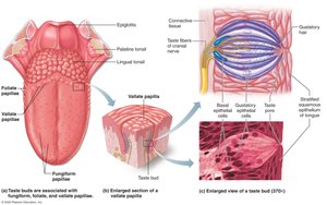
Taste Sensation and the Gustatory Pathway
There are five basic taste qualities: sweet, sour, salty, bitter, and umami (elicited by glutamate). Contrary to the traditional 'taste map,' all taste modalities can be detected from all areas containing taste buds.
Gustatory pathway: Taste information is transmitted to the cerebral cortex primarily via the facial (VII) and glossopharyngeal (IX) nerves, with some input from the vagus (X) nerve. Sensory neurons synapse in the solitary nucleus of the medulla, then relay impulses to the thalamus and ultimately to the gustatory area of the cerebral cortex in the insula.
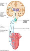
Smell (Olfaction)
Smell, or olfaction, is mediated by olfactory receptors located in the olfactory epithelium, a pseudostratified columnar tissue in the nasal cavity. The olfactory epithelium contains three main cell types: olfactory sensory neurons, supporting epithelial cells, and olfactory stem cells.
Olfactory sensory neurons: Have apical dendrites ending in a knob, from which olfactory cilia radiate. These cilia act as receptive structures for odor molecules, which must be dissolved in mucus to be detected.
Pathway: Axons of olfactory sensory neurons bundle to form filaments of the olfactory nerve, which penetrate the cribriform plate of the ethmoid bone and synapse with mitral cells in the olfactory bulbs. Mitral cells transmit impulses along the olfactory tract to the limbic system and primary olfactory cortex.
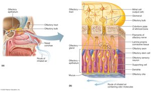
Disorders of Chemical Senses
Anosmia: Absence of the sense of smell, which may result from injury, colds, allergies, or zinc deficiency.
Uncinate fits: Distortions of smell or olfactory hallucinations, often due to irritation of olfactory pathways after brain surgery or trauma.
The Eye and Vision
General Features of Vision
Vision is the dominant sense in humans, with 70% of all sensory receptors located in the eyes and 40% of the cerebral cortex involved in processing visual information. Only the anterior one-sixth of the eye's surface is visible.
Accessory Structures of the Eye
The eye is protected and maintained by several accessory structures:
Eyebrows: Coarse hairs on the superciliary arches.
Eyelids (palpebrae): Separated by the palpebral fissure and meet at medial and lateral angles. Contain tarsal plates (connective tissue) and tarsal glands (modified sebaceous glands).
Lacrimal caruncle: Reddish elevation at the medial angle of the eye.
Conjunctiva: Transparent mucous membrane with palpebral and bulbar portions, forming the conjunctival sac.
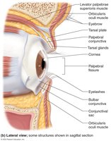
Lacrimal Apparatus
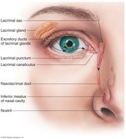
The lacrimal apparatus keeps the surface of the eye moist. The lacrimal gland produces lacrimal fluid (tears), which drains into the lacrimal sac and then empties into the nasal cavity.
Extrinsic Eye Muscles
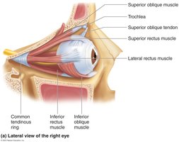
Six extrinsic eye muscles control the movement of the eyeball. These muscles originate in the walls of the orbit and insert on the outer surface of the eyeball. The annular (common tendinous) ring is the origin for four rectus muscles.
Lateral rectus
Medial rectus
Superior rectus
Inferior rectus
Superior oblique
Inferior oblique
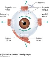
Summary Table: Extrinsic Eye Muscles, Actions, and Innervation
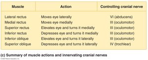
Muscle | Action | Controlling Cranial Nerve |
|---|---|---|
Lateral rectus | Moves eye laterally | VI (abducens) |
Medial rectus | Moves eye medially | III (oculomotor) |
Superior rectus | Elevates eye and turns it medially | III (oculomotor) |
Inferior rectus | Depresses eye and turns it medially | III (oculomotor) |
Inferior oblique | Elevates eye and turns it laterally | III (oculomotor) |
Superior oblique | Depresses eye and turns it laterally | IV (trochlear) |
Additional info: The coordination of these muscles allows for precise and smooth movement of the eyes, essential for binocular vision and depth perception.
