 Back
BackThe Urinary System: Structure and Function of the Nephron and Renal Processes
Study Guide - Smart Notes
 Tailored notes based on your materials, expanded with key definitions, examples, and context.
The Urinary System
Overview and Functional Importance
The urinary system is essential for maintaining the body's internal environment by regulating water, solute concentrations, and removing metabolic wastes. The kidneys are the primary organs, supported by the ureters, urinary bladder, and urethra.
Regulation of water and solute balance: Maintains osmotic concentration and ion levels in extracellular fluid.
Excretion: Removes metabolic wastes, toxins, and drugs from the body.
Endocrine functions: Produces erythropoietin (stimulates RBC production) and renin (regulates blood pressure).
Other functions: Activates vitamin D and carries out gluconeogenesis during prolonged fasting.
Nephrons: Structure and Function
General Structure of the Nephron
Nephrons are the structural and functional units of the kidney, responsible for urine formation. Each kidney contains over one million nephrons, each consisting of a renal corpuscle and a renal tubule.
Renal corpuscle: Includes the glomerulus and glomerular (Bowman's) capsule.
Renal tubule: Subdivided into the proximal convoluted tubule (PCT), nephron loop (loop of Henle), and distal convoluted tubule (DCT).
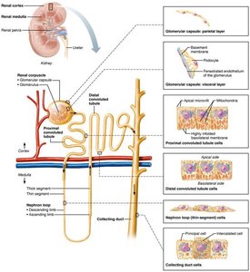
Renal Corpuscle
Glomerulus: A tuft of fenestrated capillaries specialized for filtration.
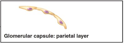
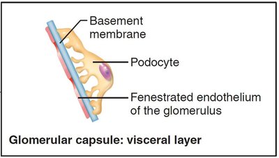
Glomerular (Bowman's) capsule: Surrounds the glomerulus and consists of two layers:
Parietal layer: Simple squamous epithelium, forms the outer wall.
Visceral layer: Composed of podocytes with foot processes that form filtration slits.
Renal Tubule Segments and Cell Types
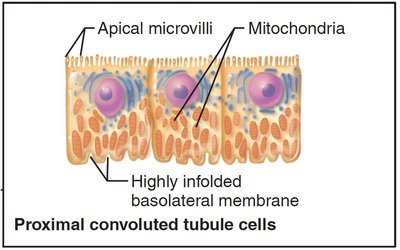
Proximal convoluted tubule (PCT): Lined with cuboidal cells with dense microvilli (brush border) and abundant mitochondria, specialized for reabsorption and secretion.
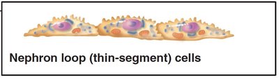
Nephron loop (Loop of Henle): U-shaped with descending and ascending limbs; thin segment lined by simple squamous cells.
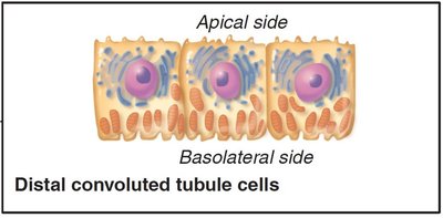
Distal convoluted tubule (DCT): Cuboidal cells with fewer microvilli, primarily involved in secretion.
Collecting duct: Contains principal cells (water and Na+ balance) and intercalated cells (acid-base balance).
Classes of Nephrons
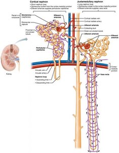
Cortical nephrons: 85% of nephrons, located almost entirely in the cortex.
Juxtamedullary nephrons: Long loops extend deep into the medulla, essential for producing concentrated urine.
Nephron Capillary Beds
Glomerulus, Peritubular Capillaries, and Vasa Recta
Each nephron is associated with two capillary beds: the glomerulus (for filtration) and peritubular capillaries (for reabsorption and secretion). Juxtamedullary nephrons also have vasa recta, which are important for urine concentration.
Juxtaglomerular Complex (JGC)
Structure and Function
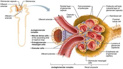
The JGC is a specialized region where the distal ascending limb of the nephron loop contacts the afferent arteriole. It regulates filtrate formation and blood pressure.
Macula densa: Chemoreceptors that sense NaCl content in filtrate.
Granular (JG) cells: Mechanoreceptors that sense blood pressure and secrete renin.
Extraglomerular mesangial cells: May relay signals between macula densa and granular cells.
Renal Processes: Filtration, Reabsorption, and Secretion
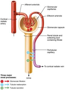
Three Major Renal Processes
Urine formation involves three key processes:
Glomerular filtration: Passive process producing cell- and protein-free filtrate.
Tubular reabsorption: Selective return of 99% of substances from filtrate to blood.
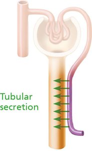
Tubular secretion: Selective movement of substances from blood to filtrate.
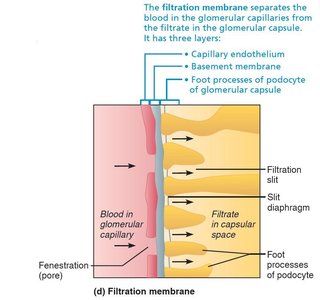
Glomerular Filtration and the Filtration Membrane
The filtration membrane consists of three layers: fenestrated endothelium, basement membrane, and podocyte foot processes. It allows passage of water and small solutes but restricts proteins and cells.
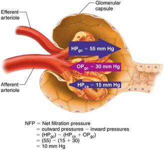
Forces Affecting Filtration
Filtration is driven by hydrostatic pressure in the glomerular capillaries and opposed by hydrostatic pressure in the capsular space and colloid osmotic pressure in the capillaries. The net filtration pressure (NFP) determines the glomerular filtration rate (GFR).
Equation for NFP:
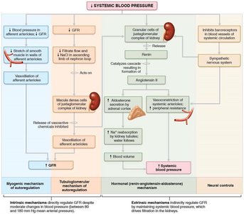
Regulation of Glomerular Filtration Rate (GFR)
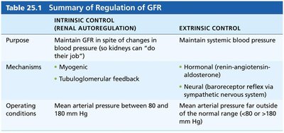
GFR is regulated by intrinsic (renal autoregulation) and extrinsic (neural and hormonal) mechanisms to maintain homeostasis and systemic blood pressure.
Intrinsic controls: Myogenic mechanism and tubuloglomerular feedback.
Extrinsic controls: Sympathetic nervous system and renin-angiotensin-aldosterone system.
Intrinsic Control (Renal Autoregulation) | Extrinsic Control |
|---|---|
Maintain GFR despite changes in blood pressure | Maintain systemic blood pressure |
Myogenic, Tubuloglomerular feedback | Hormonal (RAAS), Neural (baroreceptor reflex) |
MAP 80–180 mm Hg | MAP <80 or >180 mm Hg |
Tubular Reabsorption
Mechanisms of Reabsorption
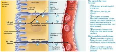
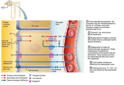
Reabsorption occurs via transcellular (through cells) and paracellular (between cells) routes. Sodium reabsorption is primarily active, driving the reabsorption of other solutes and water.
Reabsorptive Capabilities of Renal Tubules and Collecting Ducts
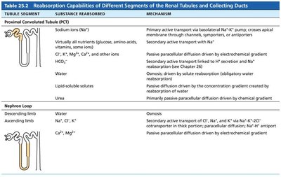
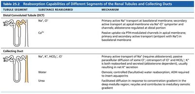
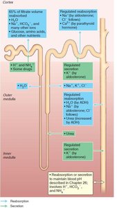
Different segments of the nephron have specialized reabsorptive functions. The PCT reabsorbs most nutrients, water, and ions; the nephron loop establishes the medullary gradient; the DCT and collecting duct are hormonally regulated.
Tubule Segment | Substance Reabsorbed | Mechanism |
|---|---|---|
Proximal Convoluted Tubule (PCT) | Na+, nutrients, Cl-, K+, HCO3-, water, urea | Active and passive transport, osmosis |
Nephron Loop | Descending: water; Ascending: Na+, Cl-, K+ | Osmosis, active and passive transport |
Distal Convoluted Tubule (DCT) | Na+, Cl-, Ca2+ | Active transport, hormone-regulated |
Collecting Duct | Na+, K+, HCO3-, Cl-, water, urea | Active and passive transport, osmosis, hormone-regulated |
Tubular Secretion
Purpose and Mechanisms
Tubular secretion removes substances from blood into the filtrate, primarily in the PCT. It is essential for eliminating drugs, excess K+, and controlling blood pH.
Regulation of Urine Concentration and Volume
Medullary Osmotic Gradient and Countercurrent Mechanisms
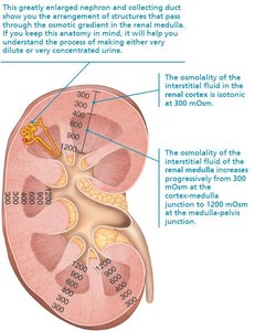
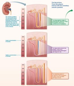
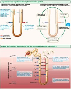
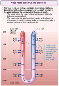
The kidneys maintain body fluid osmolality by creating a medullary osmotic gradient using countercurrent mechanisms. The nephron loop acts as a countercurrent multiplier, and the vasa recta as a countercurrent exchanger.
Formation of Dilute or Concentrated Urine
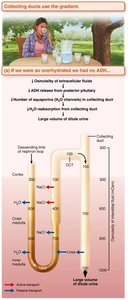
Depending on hydration status and ADH levels, the kidneys can produce dilute or concentrated urine by adjusting water reabsorption in the collecting ducts.
Renal Clearance
Definition and Clinical Application
Renal clearance is the volume of plasma cleared of a substance per unit time. It is used to assess kidney function and glomerular filtration rate (GFR).
Equation:
C: Renal clearance rate (ml/min)
U: Concentration of substance in urine (mg/ml)
V: Urine flow rate (ml/min)
P: Concentration of substance in plasma (mg/ml)
Physical and Chemical Properties of Urine
Normal and Abnormal Constituents
Normal urine: 95% water, 5% solutes (urea, uric acid, creatinine, ions)
Abnormal findings: Blood proteins, WBCs, bile pigments, or abnormal solute concentrations may indicate pathology.
Ureters, Urinary Bladder, and Urethra
Structure and Function
Ureters: Muscular tubes transporting urine from kidneys to bladder via peristalsis.
Urinary bladder: Muscular sac for temporary urine storage; contains rugae and detrusor muscle.
Urethra: Muscular tube draining urine from bladder; differs in length and function between sexes.
Micturition (Urination)
Neural Control of Micturition
Micturition involves coordinated contraction of the detrusor muscle and relaxation of internal and external urethral sphincters, regulated by autonomic and somatic nervous systems.
Reflexive urination: In infants, triggered by bladder stretch receptors.
Voluntary control: Develops with maturation of pontine control centers in the brain.
