 Back
BackThe Urinary System: Structure, Function, and Regulation
Study Guide - Smart Notes
 Tailored notes based on your materials, expanded with key definitions, examples, and context.
The Urinary System
Overview
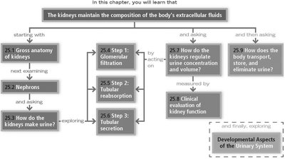
The urinary system is essential for maintaining the composition of the body's extracellular fluids by filtering blood, removing waste, and regulating water and electrolyte balance. The main organs include the kidneys, ureters, bladder, and urethra.
25.1 Gross Anatomy of the Kidneys
Location and External Anatomy
The kidneys are bean-shaped organs located retroperitoneally in the superior lumbar region. Their medial surface features a concave renal hilum, which leads into the renal sinus and serves as the entry and exit point for blood vessels, nerves, and lymphatics.
Renal fascia: Anchors the kidney and adrenal gland to surrounding structures.
Perirenal fat capsule: Cushions the kidney.
Fibrous capsule: Protects the kidney from infection.
Internal Gross Anatomy
Renal cortex: The outer region, containing most nephrons.
Renal medulla: The inner region, organized into pyramids.
Renal pelvis: Funnel-shaped tube that collects urine.
Major and minor calyces: Collect urine and empty it into the renal pelvis.
Blood and Nerve Supply
Blood enters via renal arteries and progresses through segmental, lobar, interlobar, arcuate, and cortical radiate arteries.
Afferent arterioles branch from cortical radiate arteries and supply the nephrons.
Veins trace the arterial path in reverse, draining into the renal vein.
The renal plexus regulates blood flow and nephron activity.
25.2 Nephrons: Functional Units of the Kidney
Anatomy of a Nephron
Each nephron consists of a renal corpuscle and a renal tubule. The renal corpuscle includes the glomerulus (a tuft of capillaries) and the glomerular (Bowman's) capsule.
Glomerulus: Fenestrated capillaries allow formation of filtrate.
Glomerular capsule: Parietal layer forms structure; visceral layer (podocytes) facilitates filtration.
Renal Tubule and Collecting Duct
Proximal convoluted tubule: Dense microvilli for absorption.
Nephron loop: Descending limb (permeable to water), ascending limb (permeable to ions).
Distal convoluted tubule: Few microvilli; involved in secretion and absorption.
Collecting duct: Principal cells regulate water/Na+; intercalated cells regulate acid-base balance.
Types of Nephrons
Cortical nephrons: 85%, mostly in cortex.
Juxtamedullary nephrons: 15%, near cortex-medulla junction; important for urine concentration.
Nephron Capillary Beds
Glomerulus: Specialized for filtration; high pressure maintained by afferent/efferent arterioles.
Peritubular capillaries: Low-pressure, absorb solutes and water.
Vasa recta: Parallel juxtamedullary nephrons; maintain medullary osmotic gradient.
Juxtaglomerular Complex (JGC)
Macula densa: Chemoreceptors monitoring NaCl in filtrate.
Granular cells: Mechanoreceptors monitoring blood pressure; secrete renin.
25.3 Key Processes of Urine Formation
Three Major Renal Processes
Glomerular filtration: Passive process producing filtrate.
Tubular reabsorption: Selective movement of water and solutes from filtrate back to blood.
Tubular secretion: Selective movement of solutes from blood into filtrate.
About 180 L of filtrate is formed daily, but less than 1% leaves the body as urine.
25.4 Urine Formation, Step 1: Glomerular Filtration
Filtration Membrane and Pressures
Filtration membrane: Three layers—fenestrated endothelium, basement membrane, podocytes.
Net filtration pressure (NFP): Balance of hydrostatic and osmotic pressures.
Equation for Net Filtration Pressure:
HPgc: Hydrostatic pressure in glomerular capillaries (~55 mm Hg).
HPcs: Hydrostatic pressure in capsular space.
OPgc: Colloid osmotic pressure in glomerular capillaries.
Regulation of Glomerular Filtration Rate (GFR)
Renal autoregulation: Myogenic and tubuloglomerular feedback mechanisms.
Extrinsic controls: Sympathetic nervous system and renin-angiotensin-aldosterone mechanism.
25.5 Urine Formation, Step 2: Tubular Reabsorption
Mechanisms of Reabsorption
Active reabsorption: Requires ATP; mainly for Na+.
Passive reabsorption: Diffusion, facilitated diffusion, osmosis.
Most nutrients (glucose, amino acids) are reabsorbed in the proximal convoluted tubule. Water reabsorption in the collecting duct is regulated by antidiuretic hormone (ADH).
Hormonal Regulation
ADH: Increases water reabsorption, concentrates urine.
Aldosterone: Promotes Na+ reabsorption, dilutes urine.
Atrial natriuretic peptide (ANP): Promotes Na+ loss, concentrates urine.
25.6 Urine Formation, Step 3: Tubular Secretion
Importance of Tubular Secretion
Disposes of unwanted solutes.
Eliminates reabsorbed solutes.
Rids the body of excess K+.
Controls blood pH.
25.7 Regulation of Urine Concentration and Volume
Medullary Osmotic Gradient
Countercurrent multiplier: Creates gradient via ion and water absorption in nephron loop.
Countercurrent exchanger: Maintains gradient by vasa recta.
Urea recycling: Contributes to medullary osmotic gradient.
ADH release increases water reabsorption, forming concentrated urine. Diuretics increase urine output by inhibiting Na+ reabsorption.
25.8 Clinical Evaluation of Kidney Function
Renal Clearance
Volume of plasma cleared of a substance per unit time.
Inulin is used as a standard for GFR measurement.
Clearance values indicate whether a substance is reabsorbed, secreted, or neither.
Physical and Chemical Properties of Urine
Fresh urine is clear, pale to deep yellow, slightly aromatic.
pH is usually around 6, but can range from 4.5–8.0.
Urine is 95% water, 5% solutes (mainly urea, creatinine, uric acid).
Abnormal Urinary Constituents
Glucose: Glycosuria (diabetes mellitus).
Proteins: Proteinuria (renal disease).
Blood: Hematuria (trauma, infection).
Bile pigments: Bilirubinuria (liver disease).
Ketones: Ketonuria (starvation, diabetes).
25.9 Transport, Storage, and Elimination of Urine
Ureters
Convey urine from kidneys to bladder.
Wall consists of mucosa, muscularis, and adventitia.
Urinary Bladder
Muscular sac for urine storage.
Located retroperitoneally on pelvic floor.
Wall has adventitia, detrusor muscle, and mucosa.
Capacity ~500 ml; expands as it fills.
Urethra
Drains urine from bladder.
Shorter in females (3–4 cm) than males (~20 cm).
Internal sphincter (involuntary), external sphincter (voluntary).
Micturition (Urination)
Requires detrusor contraction, internal sphincter opening, external sphincter opening.
Controlled by spinal reflexes and centers in the pons.
Developmental Aspects of the Urinary System
Embryonic Development
Three sets of kidneys: pronephros, mesonephros, metanephros.
Metanephros forms the definitive kidney.
Cloaca subdivides to form rectum, anal canal, urogenital sinus (bladder, urethra).
Postnatal and Aging Changes
Newborns void frequently; kidneys mature by two months.
Voluntary control of sphincters develops by age 4.
Kidney function declines with age; bladder shrinks and loses tone.
Recent Discoveries and Research Topics
Artificial kidneys and organoids for disease study.
Kidney disease increases risk of diabetes.
Effects of drugs, hypertension, shock, kidney stones, dialysis, and incontinence treatments on kidney function.
