 Back
BackThe Urinary System: Structure, Function, and Physiology
Study Guide - Smart Notes
 Tailored notes based on your materials, expanded with key definitions, examples, and context.
The Urinary System
Introduction to the Urinary System
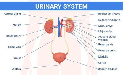
The urinary system is essential for removing metabolic wastes, regulating fluid and electrolyte balance, and maintaining homeostasis. It consists of the kidneys, ureters, urinary bladder, and urethra, each playing a specific role in urine formation and elimination.
Excretion: Removal of metabolic wastes from body fluids.
Elimination: Discharge of wastes from the body.
Homeostatic Regulation: Maintains volume and solute concentration of blood.
Urinalysis and Clinical Relevance
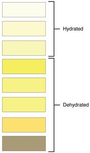
Doctors often request urine samples to screen for diseases, detect infections, and diagnose medical conditions. The color and composition of urine can provide important diagnostic information.
Urochrome: The yellow pigment in urine, derived from the breakdown of hemoglobin.
Urine Color: Indicates hydration status; darker urine suggests dehydration.
Functions of the Urinary System
The urinary system performs several vital functions:
Removal of wastes: Kidneys filter blood to remove urea, creatinine, and uric acid.
Regulation of water and electrolytes: Maintains osmotic balance and blood pressure.
Regulation of pH: Controls acid-base balance by excreting hydrogen ions and reabsorbing bicarbonate.
Organs of the Urinary System
The urinary system is composed of the following organs:
Kidneys: Paired organs that produce urine.
Ureters: Tubes that transport urine from kidneys to the bladder.
Urinary Bladder: Muscular sac for temporary urine storage.
Urethra: Tube that carries urine out of the body.
Kidney Anatomy and Histology
Location and Structure of the Kidneys
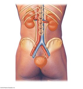
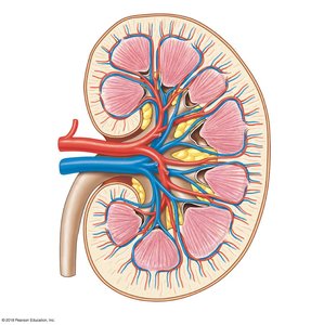
The kidneys are retroperitoneal organs located on either side of the vertebral column. The left kidney is slightly superior to the right. Each kidney is capped by an adrenal gland and stabilized by connective tissue layers.
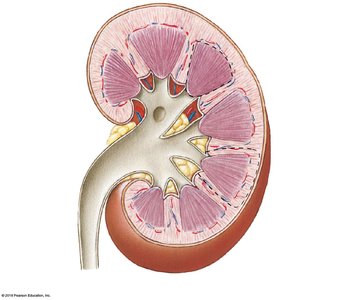
Renal Cortex: Outer region, granular appearance.
Renal Medulla: Inner region, contains renal pyramids.
Renal Sinus: Internal cavity housing renal pelvis, calyces, vessels, and nerves.
Connective Tissue Layers of the Kidney
Fibrous Capsule: Collagen fibers covering the kidney.
Perinephric Fat: Adipose tissue surrounding the capsule.
Renal Fascia: Dense outer layer anchoring the kidney.
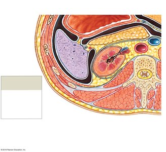
Internal Anatomy: Renal Pyramids, Columns, and Lobes
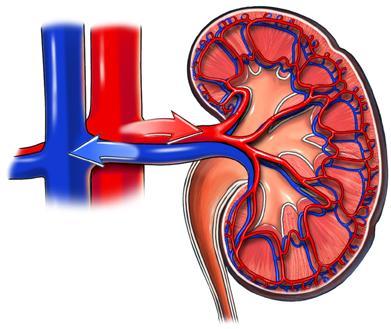
Renal pyramids are triangular structures in the medulla, separated by renal columns. Each kidney lobe consists of a pyramid, overlying cortex, and adjacent columns, and is responsible for urine production.
Renal Blood Supply
Each kidney receives blood from the renal artery, which branches into segmental, interlobar, arcuate, and cortical radiate arteries. Blood is filtered in the nephrons and returns via veins to the inferior vena cava.
Nephron Structure and Function
The Nephron: Functional Unit of the Kidney
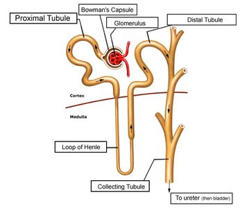
Each kidney contains about 1 million nephrons, which are responsible for filtering blood and forming urine. Nephrons consist of a renal corpuscle and a renal tubule.
Renal Corpuscle: Includes the glomerulus and Bowman’s capsule; site of filtration.
Renal Tubule: Includes the proximal convoluted tubule (PCT), nephron loop (loop of Henle), and distal convoluted tubule (DCT).
Filtration, Reabsorption, and Secretion
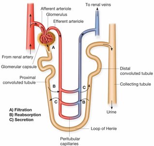
Nephrons perform three main processes:
Filtration: Blood pressure forces water and solutes from glomerulus into Bowman’s capsule.
Reabsorption: Useful substances (water, glucose, ions) are reabsorbed from the filtrate back into the blood.
Secretion: Additional wastes are secreted from blood into the tubule for excretion.
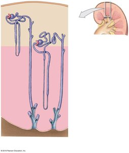
Types of Nephrons
Cortical Nephrons: 85% of nephrons; short loops of Henle, located mostly in the cortex.
Juxtamedullary Nephrons: 15% of nephrons; long loops extend deep into the medulla, important for concentrating urine.
Renal Physiology
Urine Formation
The goal of urine production is to maintain homeostasis by regulating blood volume and composition. The three main metabolic wastes are urea, creatinine, and uric acid.
Filtration: Occurs in the renal corpuscle; driven by hydrostatic pressure.
Reabsorption: Movement of water and solutes from filtrate back to blood.
Secretion: Transport of solutes from blood into the tubular fluid.
Glomerular Filtration
Filtration occurs across the filtration membrane, which consists of fenestrated endothelium, basement membrane, and podocyte foot processes. The net filtration pressure (NFP) is determined by the balance of hydrostatic and osmotic pressures.
Glomerular Hydrostatic Pressure (GHP): Pushes water and solutes out of blood.
Capsular Hydrostatic Pressure (CsHP): Opposes GHP.
Blood Colloid Osmotic Pressure (BCOP): Draws water back into blood.
Net Filtration Pressure (NFP):
Glomerular Filtration Rate (GFR): The amount of filtrate produced per minute, averaging 125 mL/min.
Hormonal Regulation of GFR
Renin-Angiotensin-Aldosterone System (RAAS): Increases blood pressure and volume, restoring GFR.
Natriuretic Peptides (ANP, BNP): Released by the heart in response to increased blood volume; increase GFR and urine output.
Reabsorption and Secretion Along the Nephron
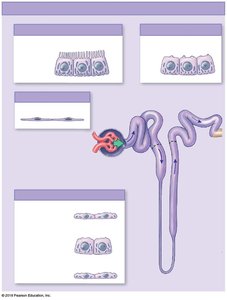
PCT: Reabsorbs water, ions, and all organic nutrients.
Nephron Loop: Descending limb reabsorbs water; ascending limb reabsorbs sodium and chloride.
DCT: Secretes ions, acids, drugs, and toxins; variable reabsorption of water and ions under hormonal control.
Collecting Duct: Variable reabsorption and secretion of water, sodium, potassium, hydrogen, and bicarbonate ions.
Countercurrent Multiplication
This process occurs in the nephron loop and is essential for concentrating urine. It involves the exchange of substances between the descending and ascending limbs, creating an osmotic gradient in the medulla.
Descending Limb: Permeable to water, not solutes.
Ascending Limb: Impermeable to water, actively transports Na+ and Cl- out.
Urine Transport, Storage, and Elimination
Ureters, Bladder, and Urethra
Urine is transported from the kidneys to the bladder via the ureters, stored in the bladder, and eliminated through the urethra. All structures are lined by transitional epithelium, allowing for distention.
Ureters: Muscular tubes with peristaltic contractions moving urine to the bladder.
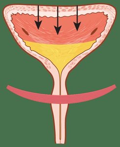
Urinary Bladder: Hollow organ with rugae for expansion; can hold up to 1 liter of urine.
Urethra: Conducts urine out of the body; contains an internal and external sphincter for control.
Micturition Reflex
The micturition reflex controls urination. Stretch receptors in the bladder wall trigger the reflex when the bladder is full, leading to contraction of the detrusor muscle and relaxation of the sphincters.
External Urethral Sphincter: Under voluntary control; relaxation allows urination.
Catheterization: Insertion of a tube to drain urine when voluntary control is lost or impaired.
Disorders of the Urinary System
Common Disorders
Kidney Stones: Crystallized minerals that can block urine flow and cause pain.
Cystitis (UTI): Infection of the bladder, more common in women due to a shorter urethra.
Bladder Cancer: Increased risk with smoking; may require chemotherapy or bladder removal.
Prostate Enlargement (BPH): Can obstruct urine flow in men.
Incontinence: Loss of voluntary control over urination.
Duplicated Ureter: Congenital condition with two ureters draining one kidney.
Renal Failure and Treatment
Dialysis: Artificial filtration of blood when kidneys fail.
Kidney Transplant: Replacement of a failed kidney with a donor organ.
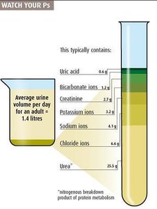
Summary Table: General Characteristics of Normal Urine
Characteristic | Normal Value |
|---|---|
Volume (per day) | 1.4 L |
Color | Pale yellow to deep amber |
pH | 4.5–8.0 (average 6.0) |
Specific Gravity | 1.003–1.030 |
Urea | 25.5 g/L |
Chloride ions | 6.6 g/L |
Sodium ions | 4.1 g/L |
Potassium ions | 2.0 g/L |
Creatinine | 1.7 g/L |
Bicarbonate ions | 1.2 g/L |
Uric acid | 0.6 g/L |
Additional info: This guide covers the structure, function, and physiology of the urinary system, including clinical relevance and common disorders, suitable for college-level Anatomy & Physiology students.
