 Back
BackThe Urinary System: Structure, Function, and Physiology
Study Guide - Smart Notes
 Tailored notes based on your materials, expanded with key definitions, examples, and context.
The Urinary System
Overview of Urinary System Structures
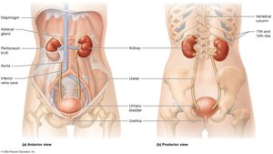
The urinary system is essential for filtering blood, removing waste, and maintaining homeostasis. It consists of paired kidneys and the urinary tract, which includes the ureters, urinary bladder, and urethra.
Kidneys: Retroperitoneal organs located against the posterior abdominal wall. The left kidney extends from T12 to L3, while the right sits lower due to the liver. Both are partially protected by the 11th and 12th ribs and are capped by adrenal glands.
Urinary Tract: Urine leaves the kidneys via ureters, is stored in the urinary bladder, and expelled through the urethra.
Overview of Kidney Function
The kidneys perform several vital functions to maintain the body's internal environment:
Removal of Metabolic Wastes: Filters waste products from the blood.
Fluid and Electrolyte Balance: Maintains osmolarity by regulating water and electrolytes (Na+, K+, Ca2+).
Acid-Base Balance: Regulates blood pH by conserving or eliminating H+ and HCO3- ions.
Blood Pressure Regulation: Influences systemic blood pressure via blood volume control and secretion of renin.
Regulation of Erythropoiesis: Releases erythropoietin to stimulate red blood cell production.
Other Metabolic Functions: Detoxifies blood, activates vitamin D, and performs gluconeogenesis.
Kidney Anatomy
External Anatomy of the Kidneys
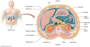
Each kidney is protected and anchored by three layers of connective tissue:
Renal Fascia: Dense irregular connective tissue anchoring the kidney.
Adipose Capsule: Middle layer of fat for shock absorption; loss can cause nephroptosis (kidney droop).
Renal Capsule: Thin, dense irregular connective tissue protecting from infection and trauma.
Internal Anatomy of the Kidneys
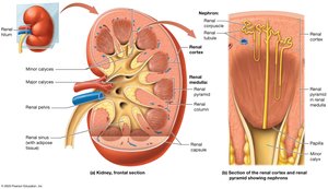
The kidney is divided into three main regions:
Renal Cortex: Outer region, rich in blood vessels.
Renal Medulla: Contains renal pyramids separated by renal columns.
Renal Pelvis: Central collecting region for urine.
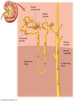
Nephrons, the functional units, are found in the cortex and medulla. Urine drains from minor calyces to major calyces, then to the renal pelvis.
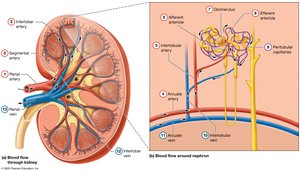
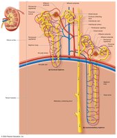
Blood Supply of the Kidneys
The kidneys receive about 25% of cardiac output. Blood flows through a series of arteries and capillaries, including the glomerulus and peritubular capillaries, before exiting via the renal vein.
Nephron Structure and Function
Microanatomy: The Nephron and Collecting System
Nephrons filter blood and modify filtrate through the renal corpuscle and renal tubule. The collecting system further processes the filtrate into urine.
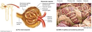
Renal Corpuscle
Glomerulus: Capillary network with fenestrations for filtration.
Bowman's Capsule: Double-layered; parietal (outer) and visceral (podocytes) layers form filtration slits.
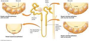
Renal Tubule
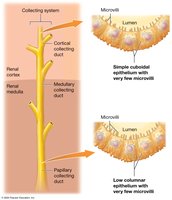
Proximal Tubule: Simple cuboidal epithelium with microvilli (brush border) for reabsorption.
Nephron Loop (Loop of Henle): Descending limb (water permeable), ascending limb (solute transport).
Distal Tubule: Simple cuboidal epithelium, less microvilli.
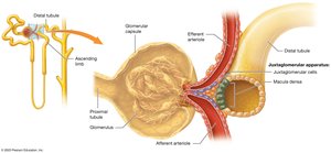
Juxtaglomerular Apparatus (JGA)
Regulates blood pressure and glomerular filtration rate via macula densa and juxtaglomerular cells.
Collecting System
Collects filtrate from distal tubules, further modifies it, and channels it to the papillary ducts as urine.
Types of Nephrons
Cortical Nephrons: 80% of nephrons, short loops, mainly in cortex.
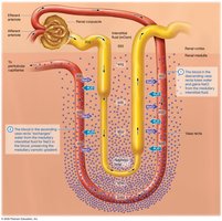
Juxtamedullary Nephrons: Long loops extend into medulla, essential for concentrating urine; surrounded by vasa recta.
Renal Physiology
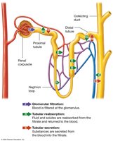
Three Basic Processes
Glomerular Filtration: Blood is filtered at the glomerulus; filtrate enters the capsular space.
Tubular Reabsorption: Substances are reclaimed from filtrate back into the blood.
Tubular Secretion: Additional substances are secreted from blood into the filtrate for excretion.
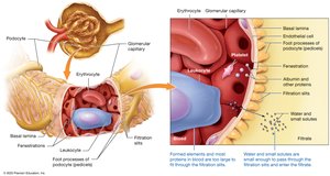
The Filtration Membrane
Fenestrated Endothelium: Allows passage of small solutes, blocks cells and platelets.
Basal Lamina: Meshwork that blocks large and negatively charged proteins.
Podocytes: Form filtration slits, allowing only small molecules (<6–7 nm) to pass.
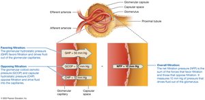
Glomerular Filtration Rate (GFR)
GFR is the rate at which filtrate is formed (about 125 mL/min). It depends on three pressures:
Glomerular Hydrostatic Pressure (GHP): Favors filtration (~50 mm Hg).
Glomerular Colloid Osmotic Pressure (GCOP): Opposes filtration (~30 mm Hg).
Capsular Hydrostatic Pressure (CHP): Opposes filtration (~10 mm Hg).
Net Filtration Pressure (NFP) is calculated as:
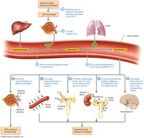
Regulation of GFR
Autoregulation: Myogenic mechanism and tubuloglomerular feedback maintain GFR within a narrow range.
Hormonal Regulation: Renin-angiotensin-aldosterone system (RAAS) and atrial natriuretic peptide (ANP) adjust GFR and blood pressure.
Neural Regulation: Sympathetic nervous system can decrease GFR during stress or blood loss.
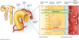
Tubular Reabsorption and Secretion
Transport Mechanisms
Paracellular Route: Substances pass between cells (e.g., ions, water).
Transcellular Route: Substances pass through cells (e.g., glucose, amino acids).
Carrier-Mediated Transport: Includes facilitated diffusion, primary and secondary active transport, antiporters, and symporters.
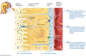
Reabsorption in the Proximal Tubule
Major Site of Reabsorption: 65% of water, most nutrients, and electrolytes are reabsorbed here.
Sodium Reabsorption: Via leak channels, symporters, and antiporters.
Glucose Reabsorption: Secondary active transport with sodium, then facilitated diffusion into blood.
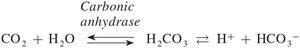
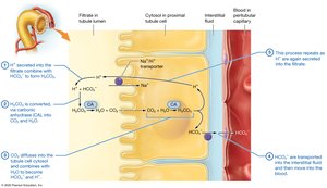
Bicarbonate Ion Reabsorption
Involves the carbonic acid-bicarbonate buffer system:
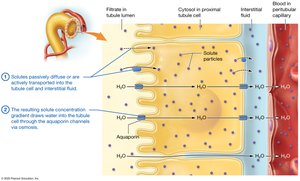
Obligatory Water Reabsorption
Water follows solutes by osmosis, aided by aquaporins, resulting in the reabsorption of about 65% of filtered water in the proximal tubule.
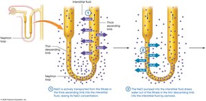
Reabsorption in the Nephron Loop
Descending Limb: Permeable to water, not solutes; filtrate becomes more concentrated.
Ascending Limb: Impermeable to water, actively transports NaCl out; filtrate becomes less concentrated.
Reabsorption and Secretion in the Distal Tubule and Collecting System
Hormone Regulation: Aldosterone increases Na+ reabsorption and K+ secretion; ADH increases water reabsorption; ANP decreases Na+ and water reabsorption.
Medullary Collecting System: Water reabsorption depends on ADH; urea recycling contributes to medullary osmotic gradient.
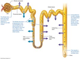
Urine Concentration and Dilution
Osmolarity of the Filtrate
Filtrate is initially isosmotic with plasma (~300 mOsm).
Descending limb concentrates filtrate; ascending limb dilutes it.
Final urine concentration is determined by facultative water reabsorption in the collecting duct, regulated by ADH.
Production of Dilute and Concentrated Urine
Dilute Urine: Produced when ADH is low; collecting ducts are impermeable to water.
Concentrated Urine: Produced when ADH is high; water is reabsorbed due to the medullary osmotic gradient.
The Countercurrent Mechanism
Countercurrent Multiplier: In juxtamedullary nephrons, establishes the medullary osmotic gradient by active transport of NaCl in the ascending limb and water reabsorption in the descending limb.
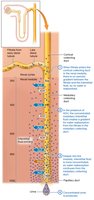
Urea Recycling: Urea diffuses into the medullary interstitium, enhancing the gradient.
Countercurrent Exchanger: Vasa recta preserves the gradient by exchanging water and solutes with the interstitium.
Urine Composition and Elimination
Urine Composition and Urinalysis
Normal Urine: Contains water, electrolytes, and metabolic wastes (urea, creatinine, uric acid).
Urinalysis: Tests for color, translucency, odor, pH, specific gravity, and abnormal solutes (e.g., blood, protein, glucose).
Renal Clearance
Definition: Rate at which kidneys remove a substance from blood; used to estimate GFR.
Creatinine: Commonly used, but not perfect due to some secretion.
Inulin: Gold standard; filtered but neither reabsorbed nor secreted.
Urine Transport, Storage, and Elimination
Urine drains from papillary ducts to minor and major calyces, then to the renal pelvis, ureters, bladder, and urethra.
Micturition: Reflex process involving bladder stretch receptors, spinal cord, and voluntary control via the external urethral sphincter.
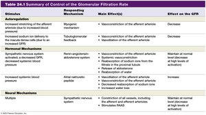
Summary Table: Control of Glomerular Filtration Rate
Stimulus | Responding Mechanism | Main Effect(s) | Effect on GFR |
|---|---|---|---|
Increased afferent arteriole stretch | Myogenic mechanism | Vasoconstriction of afferent arteriole | Decrease |
Increased NaCl at macula densa | Tubuloglomerular feedback | Vasoconstriction of afferent arteriole | Decrease |
Decreased systemic blood pressure | RAAS | Vasoconstriction of efferent arteriole, increased Na+ and water reabsorption, increased blood volume | Maintain or increase |
Increased blood volume | ANP | Dilation of afferent arteriole, constriction of efferent arteriole | Increase |
Sympathetic stimulation | Neural | Constriction of arterioles, stimulates RAAS | Decrease (high stimulation) |
Clinical Connections
Nephrolithiasis (Kidney Stones): Crystalline stones, often calcium oxalate, can cause pain and hematuria.
Glomerulonephritis: Inflammation of glomeruli, leading to proteinuria, hematuria, and possible renal failure.
Renal Failure: Acute or chronic; may require dialysis or transplantation.
Diuretics: Drugs that increase urine output by inhibiting solute reabsorption at various nephron sites.
SIADH: Excess ADH secretion causes water retention and concentrated urine.
