 Back
BackTissues: Structure, Classification, and Function in Human Anatomy
Study Guide - Smart Notes
 Tailored notes based on your materials, expanded with key definitions, examples, and context.
Tissues
Major Tissue Types
Tissues are groups of cells with similar structure and function. The four major tissue types in the human body are:
Epithelial Tissue
Connective Tissue
Muscle Tissue
Nervous Tissue
Epithelial Tissues
General Characteristics
Epithelial tissues cover body surfaces, line cavities, and form glands. They are characterized by:
Tightly packed cells with minimal extracellular matrix
Avascularity (lack of blood vessels)
Free (apical) surface exposed to the lumen or exterior
Basement membrane anchoring the epithelium to underlying connective tissue
High regenerative capacity
Innervation (presence of nerves)
Classifying Epithelial Tissues
Epithelial tissues are classified by cell shape and number of layers:
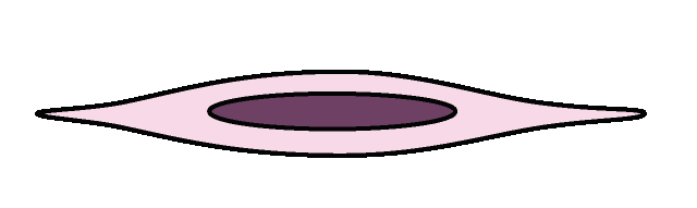
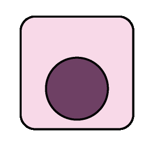
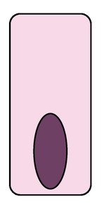
By Shape: Squamous (flat), Cuboidal (cube-shaped), Columnar (tall)
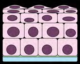
By Layers: Simple (one layer), Stratified (multiple layers)
Types of Epithelial Tissues
Simple Squamous Epithelium
Location: Alveoli of lungs, lining of blood vessels (endothelium), serous membranes (mesothelium)
Function: Rapid diffusion, filtration, and secretion
Simple Cuboidal Epithelium
Location: Kidney tubules, ducts of glands
Function: Secretion and absorption
Simple Columnar Epithelium
Location: Lining of digestive tract, gallbladder
Function: Absorption, secretion of mucus and enzymes
Specializations: Goblet cells (mucus secretion), microvilli (increase surface area)
Pseudostratified Columnar Epithelium
Location: Lining of trachea, upper respiratory tract
Function: Secretion (especially mucus), propulsion by cilia
Specialization: Cilia

Stratified Squamous Epithelium
Location: Epidermis of skin (keratinized), lining of mouth, esophagus, vagina (non-keratinized)
Function: Protection against abrasion
Keratinized: Surface cells contain keratin for waterproofing
Transitional Epithelium
Location: Urinary bladder, ureters
Function: Allows stretching and distension
Glands
Glands are composed of epithelial cells specialized for secretion. They are classified as:
Exocrine glands: Secrete products into ducts (e.g., sweat, salivary glands)
Endocrine glands: Secrete hormones directly into the bloodstream
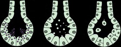
Methods of Secretion:
Merocrine: Secrete by exocytosis (e.g., sweat glands)
Apocrine: Part of cell pinches off (e.g., mammary glands)
Holocrine: Entire cell ruptures (e.g., sebaceous glands)
Connective Tissues
General Characteristics
Connective tissues support, bind, and protect other tissues and organs. They are characterized by:
Vascularity (varying degrees)
Scattered cells (e.g., fibroblasts, chondrocytes, osteocytes, macrophages, mast cells)
Extracellular matrix composed of ground substance and fibers (collagen, elastic, reticular)
Ground substance contains hyaluronic acid, providing support and hydration.
Types of Connective Tissues
Loose Connective Tissues
Areolar: Under epithelia; cushions organs; contains fibroblasts, collagen, and elastic fibers
Adipose: Under skin, around organs; stores fat; adipocytes; energy storage and insulation
Reticular: Lymphoid organs; supports cells; reticular fibers
Dense Connective Tissues
Dense Regular: Tendons, ligaments; parallel collagen fibers; resists tension
Dense Irregular: Dermis; irregular collagen fibers; withstands tension in many directions
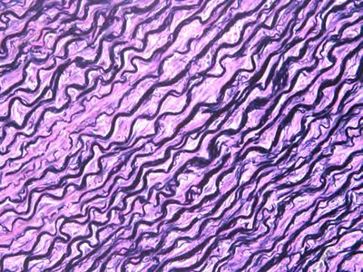
Elastic: Walls of large arteries; elastic fibers; allows recoil
Cartilage
General: Avascular, cells in lacunae, surrounded by perichondrium
Hyaline Cartilage: Ends of bones, nose, trachea; supports and reinforces; chondrocytes in matrix
Fibrocartilage: Intervertebral discs, pubic symphysis; absorbs shock; thick collagen fibers
Elastic Cartilage: External ear, epiglottis; maintains shape, flexible; elastic fibers
Osseous Tissue (Bone)
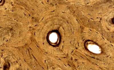
Compact Bone: Shaft of long bones; supports, protects, stores minerals; osteocytes in lacunae
Spongy Bone: Ends of long bones; produces blood cells; trabeculae structure
Fluid Connective Tissue
Blood: Within blood vessels; transports gases, nutrients, wastes; red and white blood cells in plasma
Lymph: Within lymphatic vessels; immune response; lymphocytes in fluid matrix
Muscle Tissue
General Function
Muscle tissue is specialized for contraction, producing movement. There are three types:
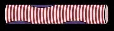
Skeletal Muscle: Attached to bones; voluntary movement; long, striated, multinucleate fibers
Cardiac Muscle: Heart wall; involuntary; striated, branched, intercalated discs
Smooth Muscle: Walls of hollow organs; involuntary; non-striated, spindle-shaped cells

Nervous Tissue
Structure and Function
Nervous tissue is found in the brain, spinal cord, and nerves. It is specialized for communication via electrical impulses.
Neurons: Conduct nerve impulses
Glial cells: Support, protect, and nourish neurons
Membranes
Body membranes are composed of epithelial and connective tissues, serving as coverings or linings.
Mucous Membrane: Lines body cavities open to the exterior (e.g., digestive, respiratory tracts); secretes mucus
Serous Membrane: Lines closed body cavities (e.g., peritoneum, pleura); secretes serous fluid
Synovial Membrane: Lines joint cavities; secretes synovial fluid for lubrication
Cutaneous Membrane: The skin; protects body surface
Tissue Repair
Repair Cells
Parenchymal cells: Functional cells of a tissue
Stromal cells: Supportive connective tissue cells
Steps of Tissue Repair
Inflammation: Damaged cells release signals; increased blood flow; degranulation of mast cells
Granulation Tissue Formation: New capillaries and fibroblasts form a temporary matrix
Maturation: Regeneration (replacement with original tissue) or fibrosis (replacement with scar tissue)
Factors Influencing Repair: Vascularity, age, metabolic rate
