 Back
BackVision: Structure and Function of the Eye and Retina
Study Guide - Smart Notes
 Tailored notes based on your materials, expanded with key definitions, examples, and context.
Special Senses: Vision
Introduction to Special Senses
The special senses include vision, taste, smell, hearing, and equilibrium. Unlike general senses, special senses utilize specialized receptor cells localized in the head region. Vision is the most studied special sense and involves complex anatomical and physiological processes within the eye and its neural pathways.
The Eye and Accessory Structures
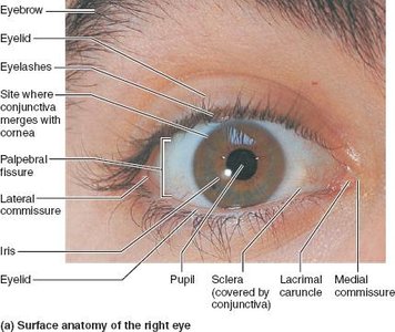
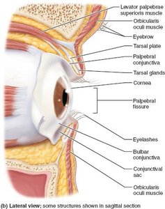
Accessory Structures of the Eye
Accessory structures protect the eye and support its function. These include the eyebrows, eyelids, conjunctiva, lacrimal apparatus, and extrinsic eye muscles.
Eyebrows: Shade the eyes and prevent sweat from reaching them.
Eyelids (palpebrae): Protect the eyes and spread secretions to keep them moist.
Conjunctiva: Transparent mucous membrane producing lubricating mucus. It has two parts: the palpebral conjunctiva (lining the eyelids) and the bulbar conjunctiva (covering the white of the eye).
Lacrimal apparatus: Produces and drains tears, keeping the eye moist and free of debris.
Extrinsic eye muscles: Six muscles that move the eyeball and help maintain its shape.
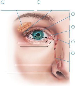
Lacrimal Apparatus
The lacrimal apparatus consists of the lacrimal gland and associated ducts. It produces tears that lubricate and protect the eye. Tears drain through the lacrimal puncta, canaliculi, sac, and finally into the nasal cavity via the nasolacrimal duct.
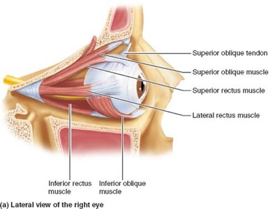
Extrinsic Eye Muscles
Six extrinsic muscles control eye movement: the superior, inferior, lateral, and medial rectus muscles, and the superior and inferior oblique muscles. These muscles originate from the bony orbit and insert on the eyeball, allowing precise movement and stabilization.
Structure of the Eyeball
Layers of the Eyeball
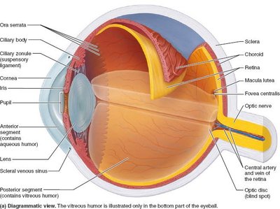
The wall of the eyeball consists of three layers:
Fibrous layer: Outermost, includes the sclera and cornea.
Vascular layer: Middle, includes the choroid, ciliary body, and iris.
Inner layer (retina): Contains photoreceptors and neurons for vision.
The lens divides the internal cavity into anterior and posterior segments, each filled with fluids called humors.
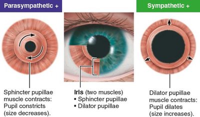
Pupil Constriction and Dilation
The iris controls the size of the pupil, regulating the amount of light entering the eye. Parasympathetic stimulation contracts the sphincter pupillae (constricting the pupil), while sympathetic stimulation contracts the dilator pupillae (dilating the pupil).
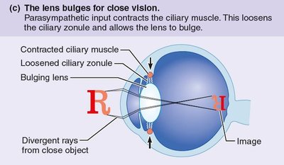
Focusing for Vision
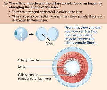
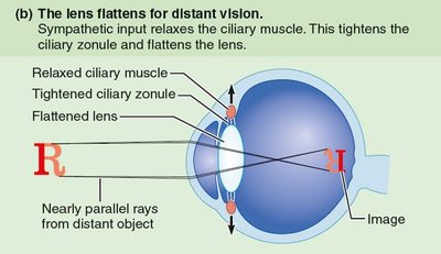
Accommodation and Lens Shape
Focusing light on the retina requires the lens to change shape, a process called accommodation. The ciliary muscle and ciliary zonule (suspensory ligament) adjust lens curvature for near or distant vision.
Distant vision: Ciliary muscle relaxes, tightening the zonule and flattening the lens.
Close vision: Ciliary muscle contracts, loosening the zonule and allowing the lens to bulge.
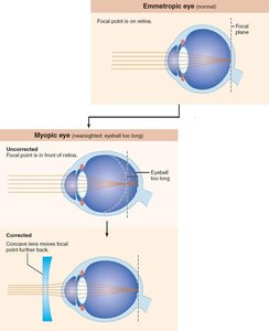
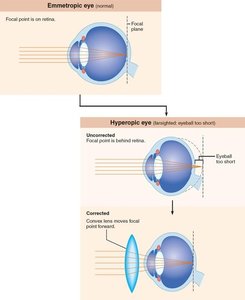
Problems of Refraction
Refraction errors occur when the eye does not focus light on the retina properly:
Myopia (nearsightedness): Eyeball too long; focal point in front of retina. Corrected with a concave lens.
Hyperopia (farsightedness): Eyeball too short; focal point behind retina. Corrected with a convex lens.
Presbyopia: Age-related loss of lens elasticity, reducing accommodation for near vision.
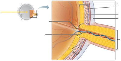
Retina: Structure and Function
Layers of the Retina
The retina consists of two main layers:
Pigmented layer: Absorbs light, prevents scattering, phagocytizes cell fragments, and stores vitamin A.
Neural layer: Contains photoreceptors (rods and cones), bipolar cells, and ganglion cells. Signals pass from photoreceptors to bipolar cells to ganglion cells, whose axons form the optic nerve.
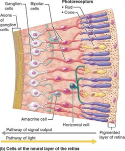
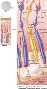
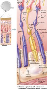
Photoreceptors: Rods and Cones
Photoreceptors are specialized neurons that detect light:
Rods: More numerous, highly sensitive to dim light, provide peripheral and night vision, but do not detect color or sharp images.
Cones: Less numerous, function in bright light, provide high-resolution color vision, concentrated in the fovea centralis.
Feature | Rods | Cones |
|---|---|---|
Vision type | Noncolor (one pigment) | Color (three pigments) |
Sensitivity | High (dim light) | Low (bright light) |
Acuity | Low (many rods per ganglion cell) | High (one cone per ganglion cell in fovea) |
Number | More numerous | Less numerous |
Location | Peripheral retina | Central retina |
Phototransduction and Information Processing
Phototransduction
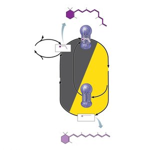
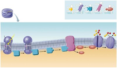
Phototransduction is the process by which light energy is converted into a graded receptor potential in photoreceptors. The key pigment in rods is rhodopsin, which consists of retinal (derived from vitamin A) and opsin. Light absorption causes retinal to change shape, triggering a cascade of events that ultimately generate electrical signals.
Formation and Breakdown of Rhodopsin
Pigment synthesis: 11-cis-retinal combines with opsin to form rhodopsin.
Pigment bleaching: Light converts 11-cis-retinal to all-trans-retinal, which detaches from opsin.
Pigment regeneration: All-trans-retinal is converted back to 11-cis-retinal in the pigmented layer, requiring ATP.
Events of Phototransduction
Light activates visual pigment (rhodopsin).
Activated pigment activates transducin (a G protein).
Transducin activates phosphodiesterase (PDE).
PDE converts cGMP to GMP, reducing cGMP levels.
cGMP-gated cation channels close, causing hyperpolarization of the photoreceptor cell.


Signal Transmission in the Retina
Photoreceptors and bipolar cells generate graded potentials. In darkness, photoreceptors release inhibitory neurotransmitter, hyperpolarizing bipolar cells. Light hyperpolarizes photoreceptors, stopping inhibitory release, allowing bipolar cells to depolarize and stimulate ganglion cells, which generate action potentials sent to the brain.
Visual Pathway to the Brain
Pathway of Visual Information
Axons of retinal ganglion cells form the optic nerve. At the optic chiasma, fibers from the medial aspect of each eye cross to the opposite side, forming optic tracts. Most fibers synapse in the lateral geniculate nucleus of the thalamus, then project to the primary visual cortex in the occipital lobe for conscious perception. Some fibers project to midbrain structures for reflexes and circadian rhythm regulation.
Depth Perception
Depth perception arises from the brain fusing slightly different images from each eye, creating a three-dimensional view. This process requires input from both eyes and is essential for accurate spatial judgment.
Light and Dark Adaptation
Adaptation Mechanisms
Light adaptation: Moving from dark to bright light causes glare as rods and cones are strongly stimulated. Rods become nonfunctional, and cones adapt quickly, improving visual acuity within minutes.
Dark adaptation: Moving from bright to dark causes temporary blindness as cones stop functioning and rods are bleached. Rhodopsin regenerates, and sensitivity increases over 20–30 minutes.
