 Back
BackVisual Tests and Common Visual Disorders: Study Notes for Anatomy & Physiology
Study Guide - Smart Notes
 Tailored notes based on your materials, expanded with key definitions, examples, and context.
Visual Tests and Experiments
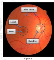
Blind Spot
The blind spot is a region on the retina known as the optic disc, where the optic nerve exits the eye and no photoreceptors are present. When an image falls on this area, it cannot be seen, creating a physiological blind spot in each eye.
Test Method: Cover one eye and focus the other on a symbol across a card; move the card until the other symbol disappears from view.
Stimulus: Visual image projected onto the optic disc.
Normal Response: The symbol disappears when it falls on the blind spot.
Accommodation
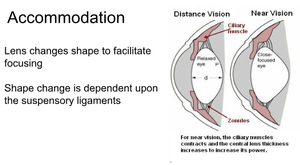
Accommodation is the process by which the eye changes the shape of the lens to focus on objects at different distances. This adjustment is controlled by the ciliary muscles and suspensory ligaments.
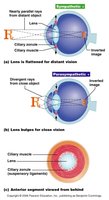
Far Vision: The lens is flat, ciliary muscles are relaxed, and suspensory ligaments are taut.
Near Vision: The lens becomes thicker and more curved as ciliary muscles contract and suspensory ligaments relax, increasing the lens's refractive power.
Test: Focus on a distant wall, then quickly shift focus to fingers held close to the face and note the sensation of the lens changing shape.
Visual Acuity
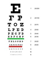
Visual acuity is the clarity or sharpness of vision, commonly measured using the Snellen chart. The results are expressed as a fraction (e.g., 20/20), where the numerator is the testing distance (in feet) and the denominator is the distance at which a person with normal vision can read the same line.
Test Method: Read letters from a Snellen chart at 20 feet, one eye at a time.
Interpretation: 20/50 means the subject sees at 20 feet what a normal eye sees at 50 feet.
Astigmatism
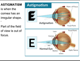
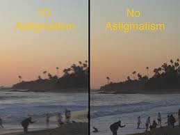
Astigmatism is a refractive error caused by an irregular curvature of the cornea or lens, resulting in blurred or distorted vision because light rays are not focused at a single point on the retina.
Test Method: Use an astigmatism chart with radial lines; if some lines appear darker or clearer, astigmatism may be present.
Symptoms: Blurred or distorted vision in parts of the visual field.
Presbyopia
Presbyopia is the age-related loss of accommodation, resulting in difficulty focusing on close objects. This occurs due to decreased elasticity of the lens with aging.
Symptoms: Difficulty reading small print or focusing on near objects, typically noticeable after age 40.
Focusing and Refraction
Focusing for Distant and Close Vision
The eye focuses light from distant and near objects by altering the shape of the lens. For distant vision, the lens is flattened; for close vision, the lens becomes more convex to increase refractive power.
Distant Vision: Light rays are nearly parallel; little lens adjustment is needed.
Close Vision: Light rays diverge; lens convexity increases via ciliary muscle contraction.
Problems of Refraction
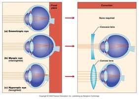
Refraction is the bending of light as it passes from one medium to another (e.g., air to cornea). Errors in refraction can lead to visual problems such as myopia (nearsightedness) and hyperopia (farsightedness).
Myopia: Light focuses in front of the retina; corrected with concave lenses.
Hyperopia: Light focuses behind the retina; corrected with convex lenses.
Color Vision and Color Blindness
Color Blindness
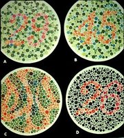
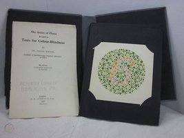
Color blindness is the inability to perceive certain colors, usually due to the absence of specific cone cells (red, green, or blue) in the retina. The Ishihara color plates are commonly used to diagnose the type and degree of color blindness.
Test Method: Identify numbers or patterns within colored dot plates.
Types: Red-green color blindness is most common.

Depth Perception
Mechanism of Depth Perception
Depth perception arises from the overlap of visual fields from both eyes, allowing the brain to fuse slightly different images into a single three-dimensional view. This is essential for judging distances and spatial relationships.
Test: Stereograms or other depth perception activities can be used to assess this ability.
Visual Reflexes
Pupillary Reflexes
The photopupillary reflex is the constriction of the pupil in response to bright light, protecting the retina from excessive illumination. The accommodation pupillary reflex occurs when focusing on a near object, causing the pupil to constrict to increase depth of focus and reduce peripheral light scatter.
Stimulus for Accommodation Reflex: Shifting focus from a distant to a near object.
Normal Response: Pupil constricts (gets smaller).
Summary Table: Common Visual Disorders
Disorder | Cause | Symptoms | Correction |
|---|---|---|---|
Myopia | Elongated eyeball or excessive curvature of cornea | Difficulty seeing distant objects | Concave lenses |
Hyperopia | Shortened eyeball or insufficient curvature of cornea | Difficulty seeing near objects | Convex lenses |
Astigmatism | Irregular curvature of cornea or lens | Blurred or distorted vision | Cylindrical lenses |
Presbyopia | Loss of lens elasticity with age | Difficulty focusing on close objects | Reading glasses |
Color Blindness | Lack of specific cone cells | Inability to distinguish certain colors | None (genetic) |
Key Equations
Snellen Visual Acuity:
Lens Equation (for reference): where is focal length, is object distance, is image distance.
