 Back
BackChapter 4 A Tour of the Cell: Structure, Function, and Microscopy
Study Guide - Smart Notes
 Tailored notes based on your materials, expanded with key definitions, examples, and context.
Chapter 4: A Tour of the Cell
Biologists Use Microscopes & Biochemistry to Study Cells
Understanding cells, the fundamental units of life, requires specialized tools and techniques. Microscopy and cell fractionation are essential for visualizing and analyzing cellular structures and functions.
Microscopy revolutionized biology in the 1600s-1700s, with Robert Hooke first observing cell walls and Antoni van Leeuwenhoek developing advanced microscopes to view 'animalcules' (microorganisms).
Microscopes allow scientists to magnify and resolve structures too small for the naked eye.

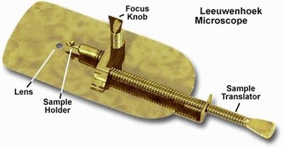
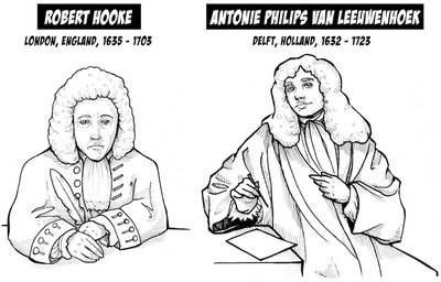
Light Microscopy (LM)
Light microscopes use visible light and glass lenses to magnify specimens, making them essential for basic cell biology.
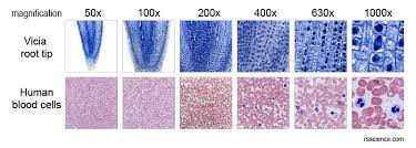
Can magnify up to ~1,000x life-size.
Enhancements in contrast and staining allow visualization of cell components.
Most organelles are too small to be resolved by LM.
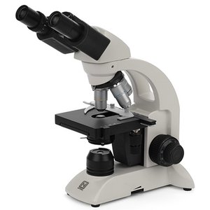
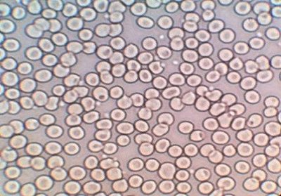
Key Parameters of Microscopy
Magnification: Ratio of image size to actual size.
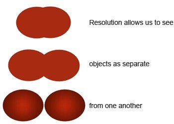
Resolution: Clarity of the image; the minimum distance two points can be distinguished as separate.
Contrast: Difference in brightness between light and dark areas.
Electron Microscopy
Electron microscopes use electron beams for much higher resolution, allowing visualization of subcellular structures.
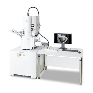
Scanning Electron Microscope (SEM): Scans the surface, producing 3D images.
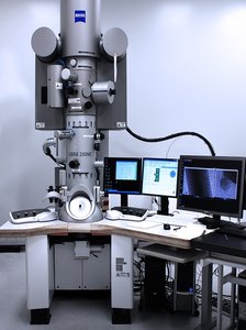
Transmission Electron Microscope (TEM): Passes electrons through specimens to study internal structures.
Cell Fractionation
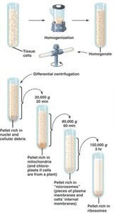
Cell fractionation separates cellular components by size and density using centrifugation, enabling the study of individual organelles and their functions.
Homogenization breaks cells apart.
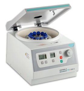
Centrifugation separates components into pellets based on size and density.
Cell Types and Their Organization
Prokaryotic vs. Eukaryotic Cells
Cells are classified as prokaryotic or eukaryotic based on structural differences.
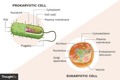
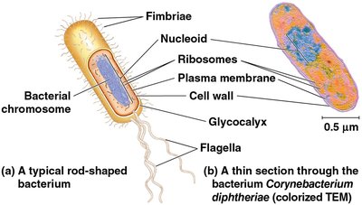
Prokaryotic cells (Bacteria, Archaea): No nucleus, DNA in nucleoid, no membrane-bound organelles, generally smaller (1-5 µm).
Eukaryotic cells (Protists, Fungi, Animals, Plants): DNA in a nucleus, membrane-bound organelles, generally larger (10-100 µm).
All cells have a plasma membrane, cytosol, chromosomes, and ribosomes.
Plasma Membrane and Cell Size
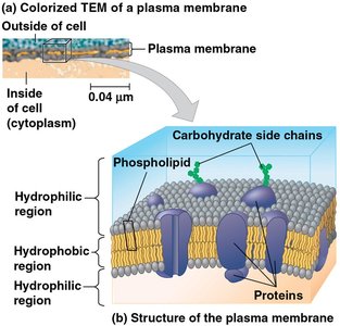
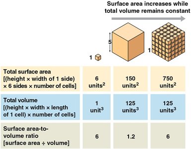
The plasma membrane is a selective barrier composed of a phospholipid bilayer. Cell size is limited by the surface area-to-volume ratio, which affects the efficiency of material exchange.
Small cells have a greater surface area relative to volume, facilitating efficient diffusion.
Internal Structures of Eukaryotic Cells
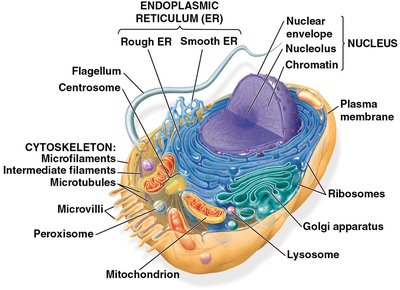
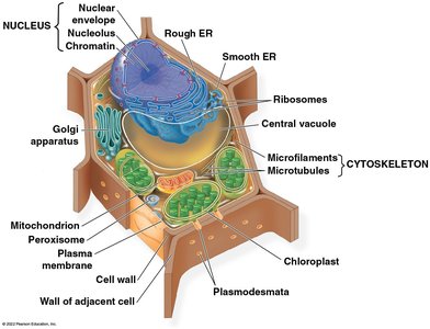
Nucleus: Genetic Control Center
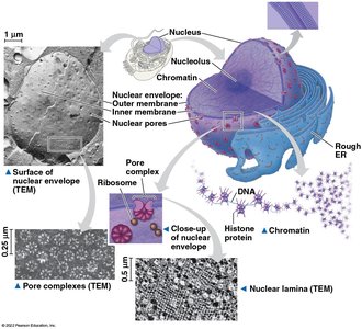
The nucleus stores genetic information and coordinates cellular activities.
Enclosed by a double-membrane nuclear envelope with pores for molecular transport.
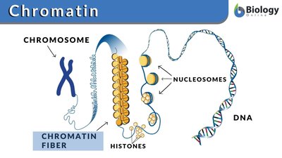
Contains DNA organized into chromosomes and chromatin (DNA + proteins).
The nucleolus is the site of ribosomal RNA (rRNA) synthesis.
Ribosomes: Protein Factories
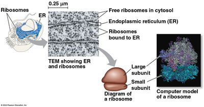
Ribosomes are complexes of rRNA and protein that synthesize proteins using genetic instructions from the nucleus.
Free ribosomes function in the cytosol; bound ribosomes are attached to the endoplasmic reticulum or nuclear envelope.
The Endomembrane System
The endomembrane system regulates protein trafficking, metabolism, and detoxification. It includes the nuclear envelope, endoplasmic reticulum (ER), Golgi apparatus, lysosomes, vacuoles, and plasma membrane.
Components are connected directly or via vesicle transfer.
Functions include protein synthesis, transport, lipid metabolism, and detoxification.
Endoplasmic Reticulum (ER)
Smooth ER: Lacks ribosomes; synthesizes lipids, metabolizes carbohydrates, detoxifies drugs/poisons, stores calcium ions.
Rough ER: Studded with ribosomes; synthesizes and secretes proteins, produces glycoproteins, distributes proteins via vesicles, and manufactures membranes.
Golgi Apparatus
Consists of flattened sacs (cisternae); modifies, sorts, and packages proteins and lipids from the ER for transport.
Lysosomes
Membranous sacs containing hydrolytic enzymes for digesting macromolecules.
Participate in phagocytosis (engulfing food particles) and autophagy (recycling cellular components).
Vacuoles
Large vesicles with diverse functions: food storage, water regulation, and enzymatic hydrolysis (especially in plants and fungi).
Central vacuole in plants stores water and ions, maintaining cell rigidity.
Energy-Transforming Organelles
Endosymbiont Theory
Mitochondria and chloroplasts originated from prokaryotic cells engulfed by ancestral eukaryotes. Both organelles have double membranes, their own DNA, and ribosomes, and replicate independently within the cell.
Mitochondria
Mitochondria are the sites of cellular respiration, converting oxygen and nutrients into ATP (energy).
Structure: Smooth outer membrane, highly folded inner membrane (cristae), intermembrane space, and mitochondrial matrix (contains DNA and ribosomes).
Chloroplasts
Chloroplasts are found in plants and algae and are the sites of photosynthesis.
Structure: Double membrane, thylakoids (stacked into grana), and stroma (internal fluid with DNA and enzymes).
Other plastids include amyloplasts (starch storage) and chromoplasts (pigment storage).
Peroxisomes
Peroxisomes are single-membrane organelles containing enzymes that detoxify harmful substances and break down fatty acids.
Oxidase converts toxins to hydrogen peroxide (H2O2), which is then converted to water and oxygen by catalase.
Cytoskeleton and Cell Structure
Cytoskeleton
The cytoskeleton is a network of protein fibers that provides structural support, organizes cell components, and enables movement.
Microtubules: Thickest fibers; maintain cell shape, guide organelle movement, and separate chromosomes during cell division.
Microfilaments (Actin): Thinnest fibers; bear tension, support cell shape, and are involved in muscle contraction and cell movement.
Intermediate Filaments: Middle diameter; reinforce cell shape and anchor organelles, more permanent than other fibers.
Motor Proteins
Motor proteins interact with the cytoskeleton to produce cell movement and transport vesicles and organelles along cytoskeletal tracks.
Extracellular Structures and Cell Junctions
Cell Walls (Plants, Fungi, Bacteria)
Plant cell walls are composed of cellulose and provide protection, structural support, and prevent excessive water uptake. Layers include the primary cell wall, middle lamella (rich in pectin), and secondary cell wall (in mature cells).
Extracellular Matrix (ECM) of Animal Cells
The ECM is a network of glycoproteins (collagen, proteoglycans, fibronectin) outside animal cells that provides structural support, cell adhesion, and communication. Integrins are membrane proteins that connect the ECM to the cytoskeleton.
Cell Junctions
Plasmodesmata: Channels in plant cell walls for transport and communication.
Tight Junctions: Seal cells together, preventing leakage (e.g., in the bladder).
Desmosomes: Anchor cells together, providing mechanical stability (e.g., in muscle and skin).
Gap Junctions: Channels for communication between animal cells (e.g., in heart muscle).
Emergent Properties of Cells
Cellular functions arise from the coordinated activity of all cellular components. The integration of organelles, membranes, and the cytoskeleton enables complex processes such as movement, metabolism, and communication.
