 Back
BackBio 100 LEC Chapter 6 Part 2
Study Guide - Smart Notes
 Tailored notes based on your materials, expanded with key definitions, examples, and context.
Bio 100 LEC Chapter 6 Part 2
Chapter 6: A Tour of the Cell
Module 5: Mitochondria, Chloroplasts, and Peroxisomes
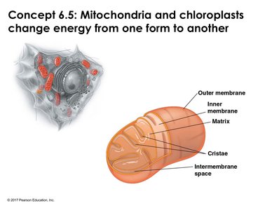
Mitochondria: Structure and Function
Mitochondria are double-membraned organelles crucial for cellular energy production, primarily generating ATP through cellular respiration. Their structure allows for compartmentalization and increased surface area, facilitating efficient metabolic processes.
Outer membrane: Defines the organelle's boundary.
Inner membrane: Highly folded into cristae, increasing surface area for metabolic reactions.
Matrix: The internal space within the folds, containing enzymes and mitochondrial DNA.
ATP generation: Most cellular events depend on mitochondria for energy.
Form and function: Cells with high energy demands (e.g., muscle cells) contain more mitochondria.
Mitochondrial DNA and Protein Synthesis
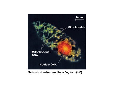
Mitochondria contain their own DNA, which is distinct from nuclear DNA. This DNA encodes some mitochondrial proteins, but most are synthesized in the cytosol and imported into the organelle.
Mitochondrial DNA: Linear chromosomes, independent from nuclear chromosomes.
Protein synthesis: Some proteins are produced by mitochondrial ribosomes; most are synthesized on free ribosomes in the cytosol.
Chloroplasts: Structure and Function
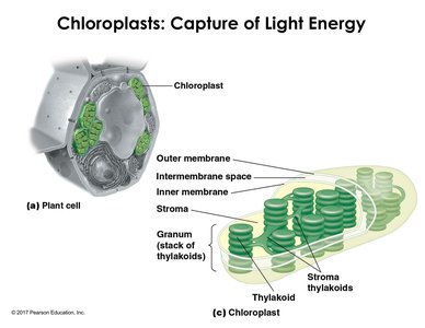
Chloroplasts are the site of photosynthesis in plants and algae. They contain the pigment chlorophyll and have a unique internal membrane system for capturing light energy.
Double membrane: Outer and inner membranes surround the organelle.
Thylakoids: Internal membrane structures arranged in stacks called grana (singular: granum).
Stroma: The fluid surrounding the thylakoids, containing enzymes for photosynthesis.
Photosynthetic function: Chloroplasts convert light energy into chemical energy.
Evolutionary Origins of Mitochondria and Chloroplasts
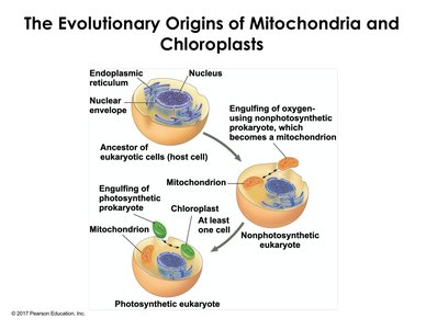
Both mitochondria and chloroplasts are believed to have originated from prokaryotic cells through endosymbiosis. This theory explains their double membranes, DNA, and ability to reproduce independently within eukaryotic cells.
Endosymbiotic theory: Eukaryotic cells engulfed prokaryotes, which became mitochondria and chloroplasts.
Mutual benefit: The engulfed cells provided energy or photosynthetic capability to the host.
Evidence: Double membranes, DNA organization, and independent proliferation.
Peroxisomes: Oxidation and Detoxification
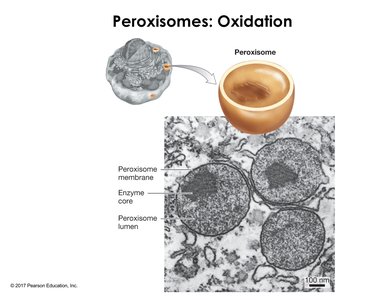
Peroxisomes are membrane-bound organelles where oxidation and reduction (redox) reactions occur. They play a key role in detoxifying harmful substances, such as hydrogen peroxide, which is converted to water and oxygen by specific enzymes.
Redox reactions: Transfer of electrons between atoms and molecules.
Detoxification: Enzymes convert hydrogen peroxide (H2O2) to water and oxygen.
Compartmentalization: Prevents damage to cellular components by isolating harmful reactions.
Module 6: The Cytoskeleton
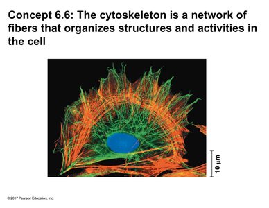
Concept 6.6: Cytoskeleton Structure and Function
The cytoskeleton is a dynamic network of fibers that organizes cell structure, anchors organelles, and facilitates cell movement. It consists of three main types of molecular structures: microtubules, microfilaments, and intermediate filaments.
Microtubules: Hollow tubes made of tubulin dimers; provide structural support and are involved in chromosome separation during cell division.
Microfilaments: Twisted chains of actin subunits; support cell shape and enable movement.
Intermediate filaments: Fibrous proteins (e.g., keratin); provide mechanical strength and maintain cell integrity.
Microtubules
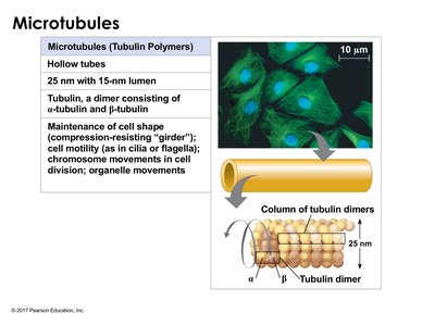
Microtubules are the largest cytoskeletal components, assembled from alpha and beta tubulin dimers. They exhibit polarity, with a faster-growing positive end, and originate from centrosomes in animal cells.
Structure: Hollow tubes, 25 nm diameter.
Subunits: Alpha and beta tubulin dimers.
Polarity: Positive and negative ends; polymerization occurs faster at the positive end.
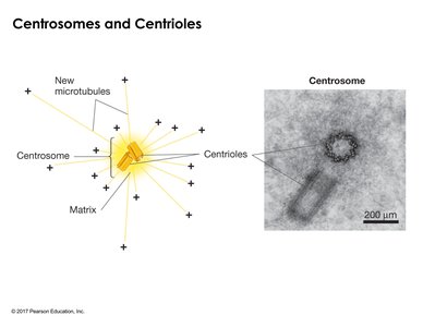
Centrosomes: Microtubule organizing centers containing centrioles arranged in nine triplets.
Functions: Structural framework, chromosome movement, organelle transport.
Motor Proteins and Microtubule Transport
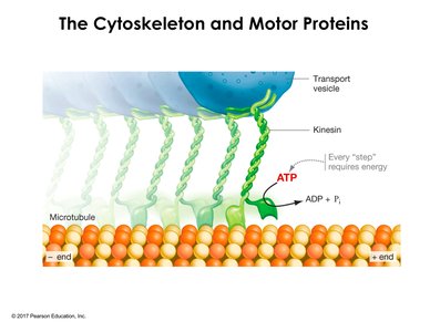
Motor proteins such as kinesin and dynein move cargo along microtubules, utilizing ATP hydrolysis for energy. Kinesin moves towards the positive end, while dynein moves towards the negative end.
Kinesin: Composed of head, stalk, and tail regions; transports vesicles.
ATP hydrolysis: Provides energy for movement.
Directionality: Kinesin moves towards the plus end; dynein towards the minus end.
Microfilaments (Actin Filaments)
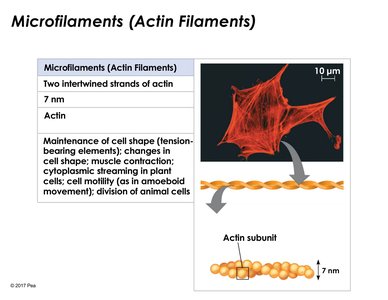
Microfilaments are solid rods about 7 nm in diameter, composed of actin subunits. They exhibit polarity and are involved in cell shape, movement, and muscle contraction.
Structure: Twisted chains of actin.
Polarity: Plus end grows faster than minus end.
Functions: Structural support, cell movement, muscle contraction, cytoplasmic streaming.
Muscle Contraction and Cell Motility
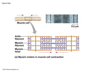
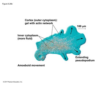
Muscle contraction is driven by interactions between actin filaments and myosin, powered by ATP hydrolysis. Actin filaments also enable cell crawling and cytoplasmic streaming in plant cells.
Myosin: Motor protein that binds actin and causes contraction.
ATP hydrolysis: Changes myosin orientation, enabling movement.
Cell crawling: Actin filaments create protrusions for movement (amoeboid movement).
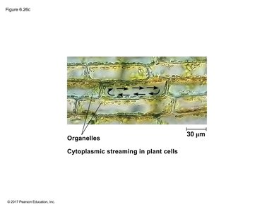
Cytoplasmic streaming: Directional flow of cytosol and organelles along actin filaments in plant cells.
Intermediate Filaments
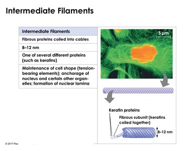
Intermediate filaments are fibrous proteins coiled into cables, providing mechanical strength and maintaining cell shape. Unlike microtubules and microfilaments, they do not exhibit polarity.
Structure: 8–12 nm diameter, composed of proteins like keratin.
Functions: Mechanical strength, anchorage of nucleus, formation of nuclear lamina.
Examples: Keratin in skin, hair, nails; nuclear lamina stabilizes nuclear envelope.
Module 7: Extracellular Components and Cell Junctions
Concept 6.7: Extracellular Components and Cellular Connections
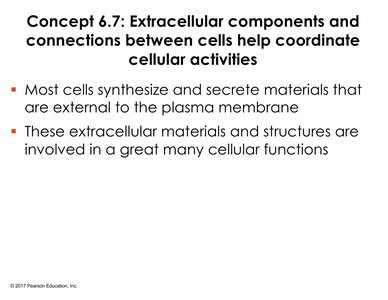
Cells synthesize and secrete materials external to the plasma membrane, forming the extracellular matrix (ECM) and cell walls. These structures coordinate cellular activities and provide structural support.
Extracellular matrix (ECM): Complex network of proteins and polysaccharides in animal cells.
Cell wall: Rigid structure in plant cells, composed of cellulose microfibrils.
Extracellular Matrix (ECM) of Animal Cells
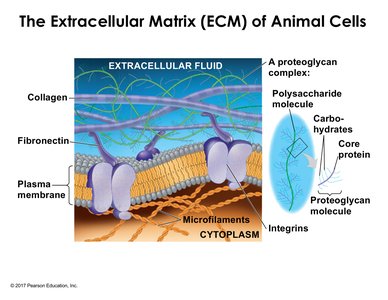
The ECM contains structural proteins (collagen), proteoglycans, adhesive glycoproteins (fibronectin), and integrins. These components provide strength, flexibility, and facilitate cell attachment and signaling.
Collagen: Imparts strength and flexibility.
Proteoglycans: Protein-polysaccharide complexes providing matrix structure.
Fibronectin: Adhesive glycoprotein binding proteoglycans and collagen.
Integrins: Cell surface receptors linking ECM and cytoskeleton.
Cell Walls of Plants
Plant cell walls provide rigidity, protection, and barriers to substance movement. They consist of cellulose microfibrils and are synthesized in stages, including the middle lamella, primary cell wall, and sometimes a secondary cell wall.
Cellulose microfibrils: Main structural component.
Middle lamella: Holds adjacent cells together.
Primary cell wall: Defines plant cell shape.
Secondary cell wall: Additional layer in mature cells.
Cell Junctions
Cell junctions are protein-based structures that allow cells to adhere, interact, and communicate. They are essential for tissue integrity and cellular coordination.
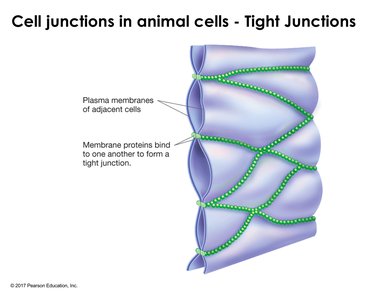
Tight junctions: Form tight seals between cells, preventing passage of substances.
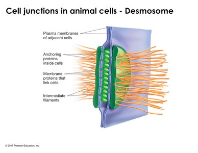
Desmosomes: Provide strong adhesion and resist mechanical stress; reinforced by intermediate filaments.
Gap junctions: Create channels for communication and movement of ions and small molecules between cells.
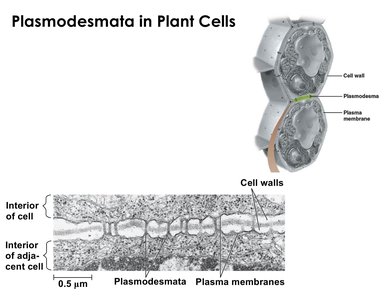
Plasmodesmata: Plant-specific junctions allowing cytoplasmic sharing between adjacent cells.
Summary Table: Cytoskeletal Components
Component | Structure | Subunits | Function |
|---|---|---|---|
Microtubules | Hollow tubes | Alpha & beta tubulin | Support, chromosome movement, organelle transport |
Microfilaments | Twisted chains | Actin | Support, movement, muscle contraction |
Intermediate Filaments | Fibrous cables | Keratin, others | Mechanical strength, nuclear lamina |
Summary Table: Cell Junctions
Junction Type | Structure | Function | Location |
|---|---|---|---|
Tight Junction | Protein seal | Barrier, restricts movement | Animal cells (e.g., intestine, bladder) |
Desmosome | Anchoring proteins, intermediate filaments | Strong adhesion, resists tension | Animal cells (e.g., skin, heart) |
Gap Junction | Protein channel | Communication, ion/molecule transfer | Animal cells (e.g., heart) |
Plasmodesmata | Membrane-lined channel | Cytoplasmic sharing | Plant cells |
Additional info: The notes expand on textbook slides with academic context, definitions, and examples to ensure completeness and clarity for exam preparation.
