 Back
BackChapter 6: A Tour of the Cell: Structure, Function, and Organization
Study Guide - Smart Notes
 Tailored notes based on your materials, expanded with key definitions, examples, and context.
Chapter 6: A Tour of the Cell
Introduction to Cell Structure and Function
The cell is the fundamental unit of structure and function in all living organisms. Cells can exist as single-celled organisms or as part of multicellular organisms, such as plants and animals. This chapter focuses on eukaryotic cells, which possess a nucleus and complex internal organization.

Microscopy and Cell Study
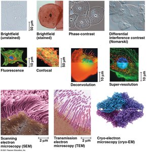
Biologists use various types of microscopes and biochemical techniques to study cells, which are typically too small to be seen with the naked eye. Understanding these tools is essential for exploring cell structure and function.
Light Microscopes (LM): Use visible light and glass lenses to magnify specimens up to 1,000 times. Key parameters include magnification, resolution, and contrast.
Electron Microscopes (EM): Use beams of electrons for higher resolution. Scanning Electron Microscopes (SEM) provide 3-D images of surfaces, while Transmission Electron Microscopes (TEM) reveal internal structures.
Advanced Techniques: Fluorescence, confocal, deconvolution, and cryo-electron microscopy enhance visualization and detail.
Cell Fractionation
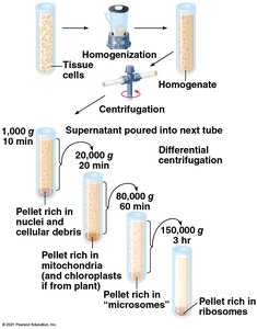
Cell fractionation is a method used to separate cellular components for study. Centrifugation allows scientists to isolate organelles and correlate their structure with function.
Differential Centrifugation: Sequentially separates cell components based on size and density.
Prokaryotic vs. Eukaryotic Cells
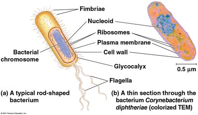
Cells are classified as prokaryotic or eukaryotic. Prokaryotic cells (Bacteria and Archaea) lack a nucleus and membrane-bound organelles, while eukaryotic cells (protists, fungi, plants, animals) have these features.
Common Features: Plasma membrane, cytosol, chromosomes, ribosomes.
Prokaryotic Cells: DNA in nucleoid, no membrane-bound organelles.
Eukaryotic Cells: DNA in nucleus, membrane-bound organelles, larger size.
Cell Size and Surface Area-to-Volume Ratio
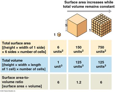
The surface area-to-volume ratio is critical for cell function, affecting the exchange of materials. As cells grow, their volume increases faster than surface area, limiting size.
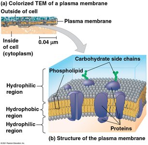
Plasma Membrane: Selective barrier for oxygen, nutrients, and waste.
Internal Organization of Eukaryotic Cells
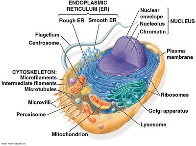
Eukaryotic cells have internal membranes that compartmentalize functions, allowing incompatible processes to occur simultaneously. The basic structure of biological membranes is a phospholipid bilayer.
The Nucleus and Ribosomes
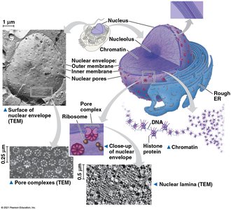
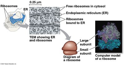
The nucleus houses genetic information and is surrounded by a double membrane (nuclear envelope). Ribosomes, composed of rRNA and protein, synthesize proteins using instructions from DNA.
Nuclear Envelope: Double membrane with nuclear pores for molecular transport.
Chromatin: DNA-protein complex that condenses into chromosomes during cell division.
Nucleolus: Site of ribosomal RNA synthesis.
Ribosomes: Free in cytosol or bound to ER/nuclear envelope.
The Endomembrane System
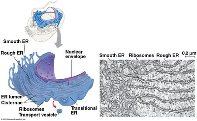
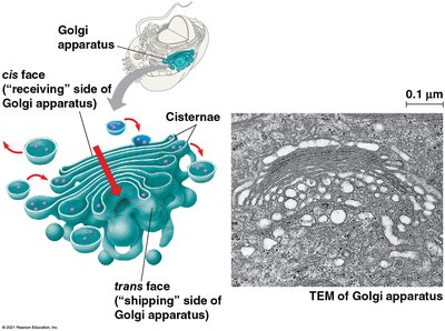
The endomembrane system regulates protein traffic and metabolic functions. It includes the nuclear envelope, endoplasmic reticulum (ER), Golgi apparatus, lysosomes, vacuoles, and plasma membrane.
Endoplasmic Reticulum (ER): Smooth ER synthesizes lipids and detoxifies; rough ER has ribosomes and produces glycoproteins.
Golgi Apparatus: Modifies, sorts, and packages ER products.
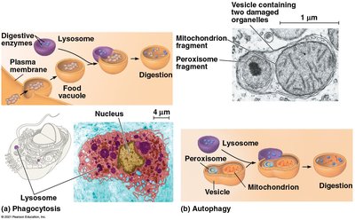
Lysosomes: Digest macromolecules and recycle cell components (autophagy).
Vacuoles: Storage and maintenance compartments; central vacuole in plants stores ions and supports growth.
Mitochondria and Chloroplasts: Energy Conversion
Mitochondria and chloroplasts are organelles that convert energy from one form to another. Mitochondria perform cellular respiration, generating ATP, while chloroplasts carry out photosynthesis.
Endosymbiont Theory: Suggests mitochondria and chloroplasts originated from engulfed prokaryotes.
Mitochondria: Double membrane, cristae, matrix; site of ATP synthesis.
Chloroplasts: Contain chlorophyll, thylakoids, stroma; site of photosynthesis.
Peroxisomes: Oxidation Reactions
Peroxisomes are single-membrane organelles containing enzymes that transfer hydrogen to oxygen, forming hydrogen peroxide. They break down fatty acids and detoxify harmful substances.
The Cytoskeleton: Support and Motility
The cytoskeleton is a network of fibers that supports cell shape, organizes organelles, and enables movement. It consists of microtubules, microfilaments (actin), and intermediate filaments.
Microtubules: Hollow rods for cell shape, organelle movement, chromosome separation.
Microfilaments: Actin filaments for cell shape, motility, and muscle contraction.
Intermediate Filaments: Provide structural stability and anchor organelles.
Extracellular Components and Cell Connections
Cells synthesize and secrete extracellular materials for protection, support, and communication.
Plant Cell Walls: Made of cellulose, provide structural support and prevent water uptake.
Extracellular Matrix (ECM) in Animals: Composed of glycoproteins, regulates cell behavior and gene activity.
Cell Junctions: Tight junctions prevent leakage, desmosomes anchor cells, gap junctions allow communication.
Plasmodesmata: Channels between plant cells for transport of water and solutes.
Integration of Cellular Components
Cellular components work together to perform complex functions. For example, macrophages coordinate cytoskeleton, lysosomes, and plasma membrane to destroy bacteria.
Summary Table: Comparison of Cytoskeletal Elements
Type | Diameter | Structure | Main Functions |
|---|---|---|---|
Microtubules | 25 nm | Hollow tubes of tubulin | Cell shape, organelle movement, chromosome separation |
Microfilaments | 7 nm | Twisted double chain of actin | Cell shape, muscle contraction, cell motility |
Intermediate Filaments | 8–12 nm | Fibrous proteins coiled into cables | Structural support, anchoring organelles |
Key Terms and Definitions
Organelle: Specialized structure within a cell performing a specific function.
Phagocytosis: Cellular process of engulfing particles.
Autophagy: Recycling of cell components by lysosomes.
Endosymbiont Theory: Hypothesis for the origin of mitochondria and chloroplasts.
Plasmodesmata: Channels connecting plant cells.
Integrins: Receptor proteins in animal cell membranes interacting with ECM.
Sample Exam Questions
Which cell structure is most likely visible with a standard light microscope? A. nucleolus
What is the likely function of a cell with a large surface area? B. rapid uptake of compounds from the environment
Why do intestinal cells not leak fluids between them? A. tight junctions bind the cells together
Additional info:
Some images and animations referenced in the original material are not included due to relevance or clarity requirements.
Tables and diagrams have been recreated and expanded for academic completeness.
