 Back
BackA Tour of the Cell: Structure, Function, and Microscopy
Study Guide - Smart Notes
 Tailored notes based on your materials, expanded with key definitions, examples, and context.
Chapter 4: A Tour of the Cell
Biologists Use Microscopes & Biochemistry to Study Cells
Understanding cells, the fundamental units of life, requires specialized tools and techniques. Microscopy and biochemical methods have enabled scientists to observe and analyze cellular structures and functions.
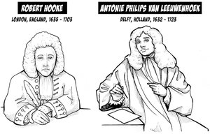
Robert Hooke: First recorded person to observe cells and cell walls.
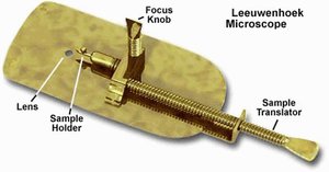
Antoni van Leeuwenhoek: Developed advanced microscopes and observed 'animalcules' (microorganisms).

Light Microscopy (LM)
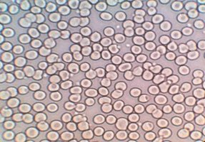
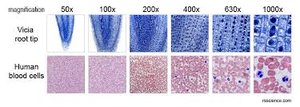
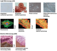
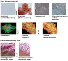
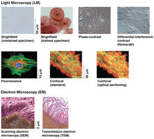
Light microscopes use visible light and glass lenses to magnify specimens, allowing observation of cells too small for the naked eye. They can achieve up to approximately 1,000x magnification.
Magnification: Ratio of image size to actual size.
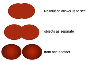
Resolution: Clarity of the image; the minimum distance two points can be distinguished as separate.
Contrast: Difference in brightness between light and dark areas.
Limitations and Advances in Microscopy
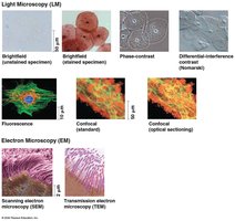
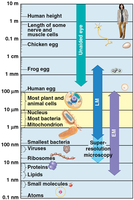
Light microscopes are limited in their ability to resolve most subcellular structures, such as organelles. Various techniques, including staining and fluorescence, enhance contrast and resolution. Recent advances, such as confocal microscopy and fluorescent labeling, have improved the ability to visualize structures as small as 10–20 µm.
Electron Microscopes
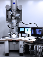
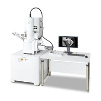
Electron microscopes use beams of electrons for much higher resolution than light microscopes, allowing detailed study of subcellular structures.
Scanning Electron Microscope (SEM): Focuses electrons on the surface, producing three-dimensional images.
Transmission Electron Microscope (TEM): Focuses electrons through a specimen, revealing internal structures.
Cell Fractionation
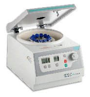
Cell fractionation is a technique that breaks cells apart and separates their components using centrifugation. This allows scientists to isolate and study individual organelles based on size and density.
Cell Types and Their Organization
Prokaryotic vs. Eukaryotic Cells
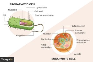
Cells are classified as prokaryotic or eukaryotic based on structural differences. Both types share certain features but differ in complexity and compartmentalization.
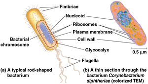
Prokaryotic Cells: Domains Bacteria and Archaea; lack a nucleus and membrane-bound organelles; DNA is in the nucleoid region; generally smaller (1–5 µm).
Eukaryotic Cells: Protists, fungi, animals, and plants; have a nucleus and membrane-bound organelles; generally larger (10–100 µm).
All cells have a plasma membrane, cytosol, chromosomes, and ribosomes.
Plasma Membrane and Cell Size
The plasma membrane is a selective barrier composed of a phospholipid bilayer, allowing passage of oxygen, nutrients, and waste. The surface area-to-volume ratio is critical for cell function; smaller cells have a greater ratio, facilitating efficient diffusion.
Internal Structures of Eukaryotic Cells
Nucleus: Location of Genetic Instructions
The nucleus contains most of the cell's DNA, organized into chromosomes. It is surrounded by a double-membrane nuclear envelope with pores for molecular transport. The nucleolus within the nucleus is the site of ribosomal RNA synthesis.
Ribosomes: Protein Factories
Ribosomes are complexes of rRNA and protein that synthesize proteins using genetic instructions. They may be free in the cytosol or bound to the endoplasmic reticulum or nuclear envelope.
The Endomembrane System
The endomembrane system regulates protein trafficking and performs metabolic functions. It includes the nuclear envelope, endoplasmic reticulum (ER), Golgi apparatus, lysosomes, vacuoles, and the plasma membrane. Components are connected directly or via vesicle transfer.
Functions: Protein synthesis, transport, metabolism of lipids, and detoxification.
Endoplasmic Reticulum (ER)
The ER is a network of membranes continuous with the nuclear envelope, comprising over half the total membrane in eukaryotic cells. It has two regions:
Smooth ER: Lacks ribosomes; synthesizes lipids, metabolizes carbohydrates, detoxifies drugs/poisons, and stores calcium ions.
Rough ER: Studded with ribosomes; synthesizes proteins and glycoproteins, distributes proteins via vesicles, and produces membranes.
Golgi Apparatus: Shipping & Receiving Center
The Golgi apparatus consists of flattened sacs (cisternae) and modifies, sorts, and packages products from the ER. It also manufactures certain macromolecules and produces glycolipids for the plasma membrane.
Lysosomes: Digestive Compartments
Lysosomes are membranous sacs containing hydrolytic enzymes for digesting macromolecules. They are involved in phagocytosis (engulfing and digesting particles) and autophagy (recycling the cell's own components).
Vacuoles: Diverse Compartments
Vacuoles are large vesicles with varied functions. In plants, the central vacuole stores water and ions. Food vacuoles form by phagocytosis, and contractile vacuoles expel excess water in protists.
Evolutionary Origins of Mitochondria & Chloroplasts
The endosymbiont theory proposes that mitochondria and chloroplasts originated as prokaryotic cells engulfed by ancestral eukaryotes. Evidence includes their double membranes, own DNA, ribosomes, and independent replication.
Mitochondria: Chemical Energy Conversion
Mitochondria are the sites of cellular respiration, generating ATP from oxygen and organic molecules. They have a double membrane, with the inner membrane folded into cristae, and contain their own DNA and ribosomes.
Chloroplasts: Site of Photosynthesis
Chloroplasts, found in plants and algae, capture light energy for photosynthesis. They contain chlorophyll, have inner and outer membranes, thylakoids (stacked into grana), and stroma (fluid with DNA and ribosomes).
Peroxisomes: Oxidative Organelles
Peroxisomes are membrane-bound organelles containing enzymes that detoxify harmful substances. Oxidase converts toxins to hydrogen peroxide, which catalase then converts to water and oxygen.
Cytoskeleton and Cell Structure
Cytoskeleton: Fiber Network Organizing Structure & Activities
The cytoskeleton is a network of protein fibers that provides structural support, maintains cell shape, and facilitates movement. It also anchors organelles and assists in intracellular transport via motor proteins.
Microtubules: Thickest fibers; shape the cell, guide organelle movement, and separate chromosomes during cell division.
Microfilaments (Actin Filaments): Thinnest fibers; bear tension, support cell shape, and are involved in muscle contraction and cell movement.
Intermediate Filaments: Medium diameter; reinforce cell shape and anchor organelles, providing mechanical stability.
Cell Walls and Extracellular Structures
Cell Walls of Plants
Plant cell walls are extracellular structures made of cellulose, providing protection, shape, and preventing excessive water uptake. Layers include the primary cell wall, middle lamella (rich in pectin), and secondary cell wall (in some cells).
Extracellular Matrix (ECM) of Animal Cells
The ECM is a network of glycoproteins (collagen, proteoglycans, fibronectin) outside animal cells, providing structural support, binding cells, and facilitating communication. Integrins are cell-surface receptors that connect the ECM to the cytoskeleton.
Cell Junctions
Types of Cell Junctions
Plasmodesmata: Channels in plant cell walls for transport of water and solutes.
Tight Junctions: Seal neighboring animal cells to prevent leakage (e.g., in the bladder).
Desmosomes: Anchor cells together, providing mechanical stability (e.g., in muscle and skin).
Gap Junctions: Channels for communication and transport between animal cells (e.g., in heart muscle).
Emergent Properties of Cells
Cellular functions arise from the coordinated activity of all cellular components. The integration of structures such as the cytoskeleton, lysosomes, and plasma membrane enables complex processes like immune responses.
