 Back
BackA Tour of the Cell – Study Notes (Campbell Biology, Chapter 6)
Study Guide - Smart Notes
 Tailored notes based on your materials, expanded with key definitions, examples, and context.
Introduction to Cells
The Cell Theory
Cells are the fundamental units of life. All living organisms are composed of cells, and all cells arise from pre-existing cells through division. Studying cells provides insight into the continuity and complexity of life, as every cell in an organism can be traced back to a single fertilized egg.
Microscopy and Cell Study
Microscopy: Tools for Studying Cells
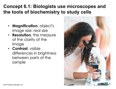
Biologists use microscopes and biochemical techniques to study cells. Microscopes are essential for revealing cellular features that are invisible to the naked eye.
Magnification: The ratio of an object's image size to its real size.
Resolution: The measure of image clarity; the ability to distinguish two close points as separate.
Contrast: The visible difference in brightness between parts of a sample.
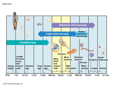
Size Range of Cells
Cells and their components vary greatly in size, from atoms and small molecules to entire cells visible to the unaided eye. Most cells require light or electron microscopy for visualization.
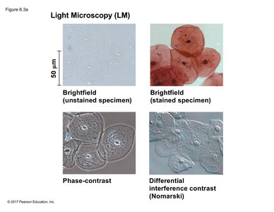
Types of Light Microscopy
Light microscopy uses visible light to illuminate specimens. Several techniques enhance visualization:
Brightfield (unstained): Minimal contrast, limited detail.
Brightfield (stained): Staining increases contrast and reveals structures like the nucleus.
Phase-contrast: Enhances contrast without staining, useful for live cells.
Differential Interference Contrast (DIC): Uses polarized light to create a 3D-like effect.
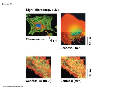
Advanced Light Microscopy
Fluorescence Microscopy: Uses fluorescent dyes or proteins to label specific cell components, which are then visualized under specific wavelengths of light.
Confocal Microscopy: Uses lasers and optical sectioning to produce sharp images of a single plane within a specimen; can be enhanced by deconvolution software for improved clarity.
Electron Microscopy
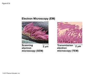
Electron microscopes use electron beams for much higher resolution than light microscopes. Two main types:
Scanning Electron Microscopy (SEM): Visualizes 3D surfaces of specimens.
Transmission Electron Microscopy (TEM): Visualizes internal structures by passing electrons through thin sections.
Cell Fractionation
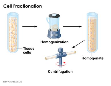
Cell fractionation separates cellular components for individual study. The process involves homogenizing tissue to release cell contents, then using centrifugation to separate components by size and density.
Differential Centrifugation
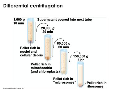
Successive centrifugation steps at increasing speeds isolate different cellular fractions:
Low speed: nuclei and debris
Medium speed: mitochondria, chloroplasts
High speed: microsomes
Very high speed: ribosomes
Cell Types and Internal Organization
Prokaryotic vs. Eukaryotic Cells
Cells are classified as prokaryotic or eukaryotic based on internal structure:
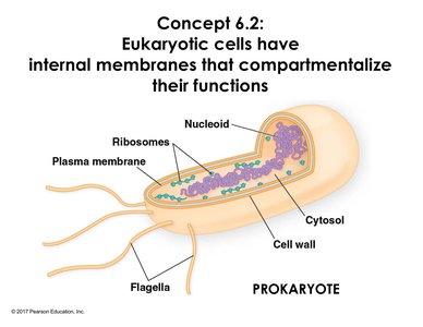
Prokaryotes: Lack a membrane-bound nucleus; DNA is in a nucleoid region. Have plasma membrane, cytosol, ribosomes, and sometimes flagella.
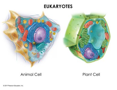
Eukaryotes: Have a nucleus with a double membrane, multiple linear chromosomes, and extensive internal compartmentalization into organelles.
The Plasma Membrane
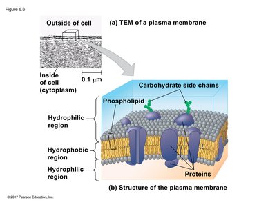
The plasma membrane is a selective barrier composed of a phospholipid bilayer with embedded proteins and carbohydrates. It regulates the passage of substances into and out of the cell and is essential for compartmentalization.
Cell Size and Surface Area-to-Volume Ratio
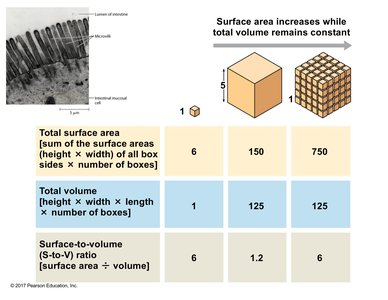
Cells are small to maximize the surface area-to-volume ratio, which is critical for efficient exchange of materials with the environment. As cells grow, volume increases faster than surface area, limiting size. Cells may have adaptations like microvilli to increase surface area.
Cube | Total Surface Area | Total Volume | Surface Area:Volume Ratio |
|---|---|---|---|
1 large | 6 | 1 | 6 |
1 medium | 150 | 125 | 1.2 |
Many small | 750 | 125 | 6 |
Genetic Material and Protein Synthesis
The Nucleus
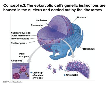
The nucleus houses chromosomes and is the site of DNA replication and transcription. It is surrounded by a double membrane (nuclear envelope) with nuclear pores for regulated exchange. The nucleolus within the nucleus assembles ribosomal RNA and ribosome subunits.
Ribosomes
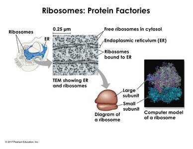
Ribosomes are macromolecular complexes of RNA and protein that synthesize polypeptides. They exist as free ribosomes in the cytosol or bound to the endoplasmic reticulum (ER). Ribosomes consist of large and small subunits that associate non-covalently.
The Endomembrane System
Components and Functions
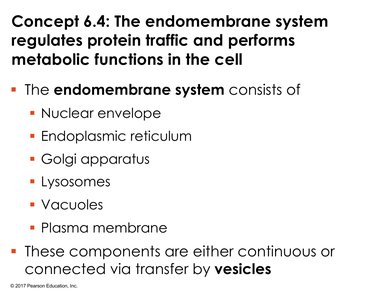
The endomembrane system regulates protein traffic and metabolic functions. It includes the nuclear envelope, endoplasmic reticulum (ER), Golgi apparatus, lysosomes, vacuoles, and plasma membrane. Components are connected directly or via vesicle transfer.
Endoplasmic Reticulum (ER)
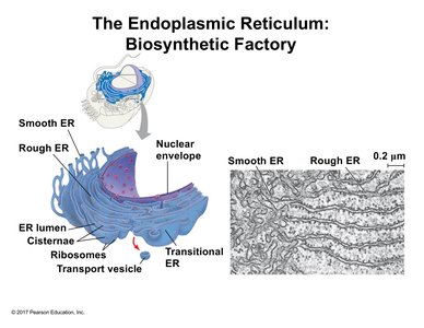
The ER is continuous with the nuclear envelope and exists in two forms:
Smooth ER: Lacks ribosomes; synthesizes lipids, detoxifies harmful substances, and stores calcium ions.
Rough ER: Studded with ribosomes; synthesizes proteins for secretion or for use in membranes and organelles. The interior is called the lumen, where proteins are folded and modified (e.g., glycosylation).
Golgi Apparatus
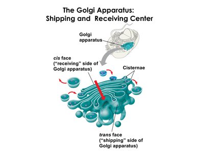
The Golgi apparatus processes, sorts, and ships proteins. It consists of flattened sacs (cisternae) with a cis face (receiving side) and a trans face (shipping side). Proteins move from the ER to the cis face, are modified, and exit via the trans face in vesicles.
Lysosomes
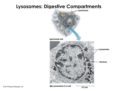
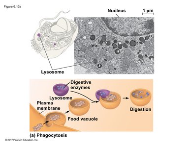
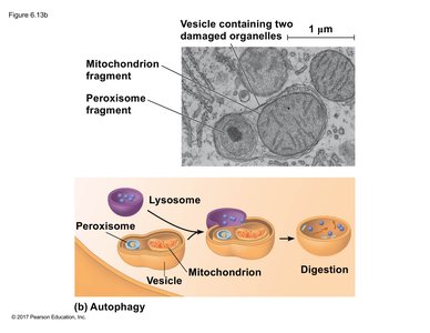
Lysosomes are membrane-bound sacs containing hydrolytic enzymes (acid hydrolases) that digest macromolecules. They function optimally in acidic conditions maintained by proton pumps. Lysosomes recycle cellular components and are involved in processes like phagocytosis and autophagy.
Vacuoles
Vacuoles are prominent in plant cells and serve as storage for water, pigments, proteins, or toxins. Some vacuoles contain hydrolases and function similarly to lysosomes in animal cells.
Energy-Transforming Organelles
Mitochondria
Mitochondria are the main site of ATP production via cellular respiration. They have a double membrane, with the inner membrane folded into cristae to increase surface area. Mitochondria contain their own DNA and ribosomes, but most proteins are imported from the cytosol.
Chloroplasts
Chloroplasts are found in plants and algae and are the site of photosynthesis. They have a double membrane and internal stacks of thylakoids (grana) containing chlorophyll. Chloroplasts also contain their own DNA and ribosomes.
Endosymbiotic Theory
Both mitochondria and chloroplasts share similarities with prokaryotes, such as double membranes, circular DNA, and independent division. The endosymbiotic theory proposes that these organelles originated from engulfed prokaryotic cells that formed a mutualistic relationship with ancestral eukaryotes.
Peroxisomes
Peroxisomes are membrane-bound organelles where oxidation-reduction (redox) reactions occur, such as the breakdown of fatty acids and detoxification of hydrogen peroxide (H2O2) into water and oxygen. Specialized enzymes (e.g., catalase) prevent cellular damage from reactive byproducts.
The Cytoskeleton
Overview
The cytoskeleton is a dynamic network of protein fibers that organizes cell structure, anchors organelles, and facilitates movement. It consists of three main types:
Microtubules: Hollow tubes made of tubulin dimers; provide structural support, serve as tracks for motor proteins, and are essential for chromosome separation during cell division.
Microfilaments (Actin Filaments): Solid rods of actin; support cell shape, enable movement (e.g., muscle contraction, cell crawling), and drive cytoplasmic streaming in plants.
Intermediate Filaments: Rope-like fibers (e.g., keratin); provide mechanical strength and maintain cell and nuclear shape. Unlike the other two, they lack polarity and are more stable.
Extracellular Components and Cell Junctions
Extracellular Matrix (ECM) in Animal Cells
The ECM is a network of proteins and carbohydrates outside animal cells that provides structural support and mediates cell signaling. Key components include:
Collagen: Provides tensile strength.
Proteoglycans: Protein-polysaccharide complexes that form a gel-like matrix.
Fibronectin: Glycoprotein that connects cells to the ECM.
Integrins: Transmembrane receptors linking the ECM to the cytoskeleton.
Cell Walls in Plants
Plant cells are surrounded by a rigid cell wall composed mainly of cellulose. The wall provides structural support, protection, and regulates water intake. The middle lamella glues adjacent cells together, and some cells develop a secondary wall for additional strength.
Cell Junctions
Tight Junctions: Seal adjacent animal cells together, preventing leakage of extracellular fluid.
Desmosomes: Anchor cells together via intermediate filaments, providing mechanical strength (abundant in tissues subject to stress, e.g., skin, heart).
Gap Junctions: Channels that allow ions and small molecules to pass directly between animal cells, enabling rapid communication (e.g., in heart muscle).
Plasmodesmata: Channels in plant cell walls that connect the cytoplasm of adjacent cells, allowing exchange of substances and communication.
