 Back
BackCell Cycle and Mitosis: Structure, Regulation, and Cancer
Study Guide - Smart Notes
 Tailored notes based on your materials, expanded with key definitions, examples, and context.
Cell Cycle and Mitosis
Key Roles of Cell Division
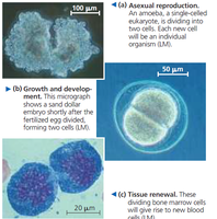
Cell division is a fundamental process in all living organisms, essential for reproduction, growth, development, and tissue renewal. The process results in the formation of genetically identical daughter cells, ensuring continuity of genetic information.
Asexual reproduction: Single-celled organisms divide to produce offspring genetically identical to the parent.
Growth and development: Multicellular organisms grow by increasing cell number through division.
Tissue renewal: Damaged or dead cells are replaced by new cells via mitosis.
Cellular Organization of Genetic Material
Genome and Chromosomes
The genome of a cell comprises all its DNA, including genes and noncoding regions. In eukaryotes, DNA is packaged into structures called chromosomes, which are highly condensed forms of chromatin (DNA plus histone proteins). Human cells contain approximately 2 meters of DNA per cell, compacted into chromosomes to fit within the nucleus.
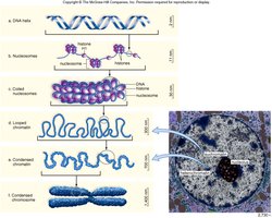
Chromatin Structure: Heterochromatin vs. Euchromatin
Heterochromatin: Densely packed, transcriptionally inactive regions of chromatin.
Euchromatin: Less condensed, transcriptionally active regions.
Chromosome Terminology
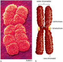
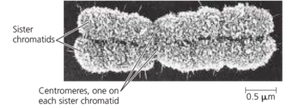
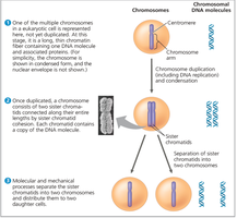
Sister chromatids: Identical copies of a chromosome connected at the centromere after DNA replication.
Centromere: Region where sister chromatids are joined and where spindle fibers attach during mitosis.
Kinetochore: Protein complex on the centromere that binds spindle microtubules.
Diploid (2n): Cells with two sets of chromosomes (e.g., somatic cells).
Haploid (n): Cells with one set of chromosomes (e.g., gametes).
Chromosome Duplication and Distribution
During cell division, chromosomes are duplicated and then distributed equally to daughter cells. This ensures each new cell receives a complete set of genetic information.
Duplicated chromosomes consist of two sister chromatids.
During mitosis, sister chromatids are separated, resulting in unduplicated chromosomes in each daughter cell.
The Cell Cycle
Overview of the Cell Cycle
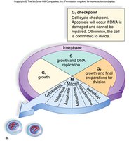
The cell cycle is a repeating series of events that lead to cell division and the production of two daughter cells. It consists of interphase (G1, S, G2) and the M phase (mitosis and cytokinesis).
G1 phase: Cell growth and metabolic activity.
S phase: DNA synthesis and replication of chromatids.
G2 phase: Further growth, protein synthesis, and preparation for division.
M phase: Includes mitosis (nuclear division) and cytokinesis (cytoplasmic division).
Interphase
G1 phase: Cell increases in size and prepares for DNA replication.
S phase: DNA is replicated, resulting in duplicated chromosomes (sister chromatids).
G2 phase: Cell continues to grow and prepares for mitosis by synthesizing proteins and organelles.
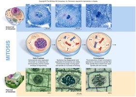
M Phase: Mitosis and Cytokinesis
Mitosis is the process of nuclear division, while cytokinesis is the division of the cytoplasm. Mitosis is divided into several stages:
Prophase: Chromatin condenses into visible chromosomes, nuclear envelope breaks down, spindle apparatus forms.
Prometaphase: Kinetochores form, spindle fibers attach to chromosomes.
Metaphase: Chromosomes align at the metaphase plate.
Anaphase: Sister chromatids are pulled apart to opposite poles.
Telophase: Nuclear envelopes reform, chromosomes decondense.
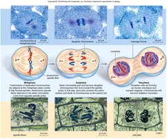
Cytokinesis
Animal cells: Cleavage furrow forms, contractile ring of microfilaments pinches the cell in two.
Plant cells: Vesicles form a cell plate that develops into a new cell wall.
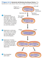
Prokaryotic Cell Division: Binary Fission
Prokaryotes, such as bacteria, divide by binary fission, a simpler process than mitosis. The single, circular DNA molecule is replicated, and the cell splits into two genetically identical daughter cells.
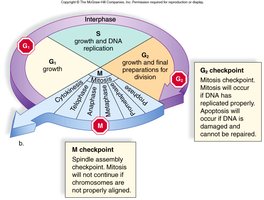
Regulation of the Cell Cycle
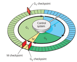
Cell Cycle Control System and Checkpoints
The cell cycle is tightly regulated by a control system with checkpoints at critical stages (G1, G2, and M). These checkpoints ensure that damaged or incomplete DNA is not passed on to daughter cells.
G1 checkpoint: Determines if the cell will proceed with division or enter a non-dividing state (G0).
G2 checkpoint: Ensures DNA replication is complete and undamaged before mitosis.
M checkpoint: Ensures all chromosomes are properly attached to the spindle before anaphase.
Cyclins and Cyclin-Dependent Kinases (Cdks)
Progression through the cell cycle is regulated by cyclins and cyclin-dependent kinases (Cdks). Cyclins are proteins whose levels fluctuate during the cell cycle, while Cdks are enzymes that, when activated by cyclins, phosphorylate target proteins to drive cell cycle events.
MPF (Maturation-Promoting Factor): A cyclin-Cdk complex that triggers the cell's passage from G2 into M phase. MPF activity peaks at metaphase and is degraded in anaphase.
External and Internal Signals
Growth factors: Proteins such as PDGF (platelet-derived growth factor) stimulate cell division.
Density-dependent inhibition: Cells stop dividing when crowded.
Anchorage dependence: Cells must be attached to a substrate to divide.
Cancer and Loss of Cell Cycle Control
What is Cancer?
Cancer is characterized by uncontrolled cell division due to the loss of normal cell cycle controls. Cancer cells exhibit abnormal behaviors, such as the formation of tumors (neoplasms), evasion of apoptosis, and the ability to metastasize (spread to distant tissues).
Benign tumors: Do not spread and are usually not life-threatening.
Malignant tumors: Invade surrounding tissues and can metastasize.
Genetic Basis of Cancer
Proto-oncogenes: Genes that promote cell division; mutations convert them to oncogenes, leading to uncontrolled growth (e.g., ras genes, BRCA1).
Tumor suppressor genes: Genes that inhibit cell division; mutations inactivate these genes, removing growth restraints (e.g., p53, RB genes).
Treatment of Cancer
Surgery: Removal of tumors.
Radiotherapy: Use of radiation to kill cancer cells.
Chemotherapy: Drugs that target rapidly dividing cells, often by interfering with mitosis (e.g., Taxol disrupts spindle formation).
Precision medicine: Genetic testing to select drugs tailored to the patient's specific mutations.
Side effects: Chemotherapy can affect normal dividing cells, leading to symptoms such as nausea and hair loss.
Summary Table: Key Differences Between Normal and Cancer Cells
Feature | Normal Cells | Cancer Cells |
|---|---|---|
Cell Cycle Control | Regulated by checkpoints | Lost or defective |
Apoptosis | Functional | Often evaded |
Growth Factors | Required for division | May divide without them |
Density-Dependent Inhibition | Present | Absent |
Anchorage Dependence | Present | Absent |
Metastasis | No | Yes (malignant) |
