 Back
BackCell Cycle, Chromosome Structure, and Mitosis: Study Notes for BIOL 1001
Study Guide - Smart Notes
 Tailored notes based on your materials, expanded with key definitions, examples, and context.
Cell Structure and Function
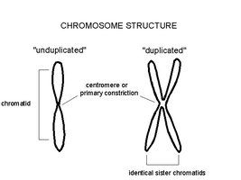
Chromosome Structure
Chromosomes are highly organized structures composed of DNA and proteins, essential for the storage and transmission of genetic information. The DNA is tightly packed to fit within the cell nucleus, and this packing is achieved through several hierarchical levels.
Chromatid: Each chromosome consists of one or two chromatids, depending on the stage of the cell cycle.
Centromere: The primary constriction point that holds sister chromatids together.
Unduplicated vs. Duplicated Chromosomes: An unduplicated chromosome has a single chromatid, while a duplicated chromosome has two identical sister chromatids joined at the centromere.
Chromatin: The complex of DNA and proteins (mainly histones) that forms chromosomes.
Example: During cell division, chromosomes duplicate to form sister chromatids, which are separated into daughter cells.
Chromosome Packing
DNA is packaged into chromosomes through a series of structural levels, allowing the long DNA molecules to fit inside the nucleus.
Double Helix: The basic structure of DNA.
Histones: Proteins around which DNA winds, forming nucleosomes.
Nucleosome: The "beads-on-a-string" form of chromatin.
Chromatin Fiber: Nucleosomes are further packed into thicker fibers.
Chromosome: The most condensed form, visible during cell division.
Example: Each DNA molecule is packaged into a chromosome that is thousands of times shorter than its extended length.

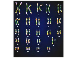
Human Karyotype
A karyotype is a visual representation of all the chromosomes in a cell, arranged in pairs. Humans have 23 pairs of chromosomes: 22 pairs of autosomes and 1 pair of sex chromosomes.
Autosomes: Chromosomes 1-22, not involved in sex determination.
Sex Chromosomes: Pair 23, determines biological sex (XX for female, XY for male).
Example: Karyotyping is used to detect chromosomal abnormalities such as Down syndrome (trisomy 21).
Cell Reproduction
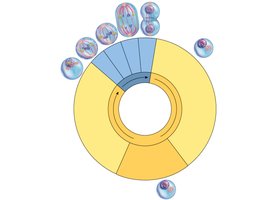
The Cell Cycle
The cell cycle is the series of events that cells go through as they grow and divide. It consists of interphase (G1, S, G2) and mitotic phase (mitosis and cytokinesis).
G1 Phase: Cell growth and differentiation.
S Phase: DNA synthesis; chromosomes are duplicated.
G2 Phase: Further growth and preparation for cell division.
Mitosis: Division of the nucleus.
Cytokinesis: Division of the cytoplasm.
Example: Most cells spend the majority of their time in interphase, preparing for division.
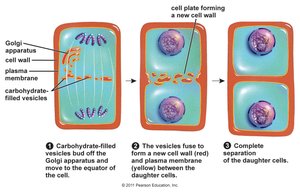
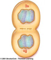
Cytokinesis in Plant Cells
Cytokinesis is the process by which the cytoplasm divides, forming two daughter cells. In plant cells, this involves the formation of a cell plate.
Cell Plate Formation: Vesicles from the Golgi apparatus fuse at the center of the cell, forming a new cell wall.
Separation: The cell plate expands outward, eventually separating the two daughter cells.
Example: Plant cells form a cell plate during cytokinesis, unlike animal cells which form a cleavage furrow.
Stages of Mitosis
Mitosis is the process by which a cell divides its nucleus and distributes identical genetic material to two daughter cells. The stages include prophase, metaphase, anaphase, and telophase.
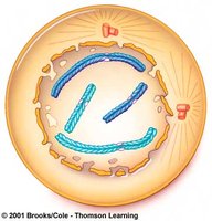
Prophase: Chromosomes condense and become visible; spindle fibers form.
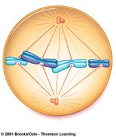
Metaphase: Chromosomes align at the cell's equator.
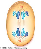
Anaphase: Sister chromatids are pulled apart to opposite poles.
Telophase: Chromosomes decondense; nuclear envelopes reform.
Example: Mitosis ensures that each daughter cell receives an identical set of chromosomes.
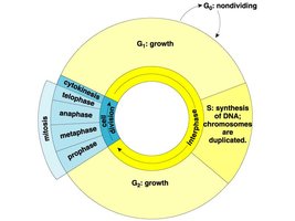
Interphase
Interphase is the longest part of the cell cycle, representing about 90% of the complete cycle. It is characterized by high metabolic activity, cell growth, and duplication of organelles and chromosomes.
Cell Mass Increase: The cell grows in size and prepares for division.
Organelle Duplication: Organelles are duplicated to ensure each daughter cell receives a full complement.
Chromosome Duplication: DNA is replicated during the S phase.
Example: Cells in interphase are not actively dividing but are preparing for the next division.

Early and Late Prophase
Prophase marks the beginning of mitosis, where chromosomes condense and spindle fibers begin to form. In late prophase, microtubules assemble, centrioles move to opposite poles, and the nuclear envelope breaks down.
Early Prophase: Chromosomes condense and become visible.
Late Prophase: Spindle fibers attach to chromosomes; nuclear envelope disintegrates.
Example: The transition from early to late prophase is critical for proper chromosome alignment and segregation.
Metaphase
During metaphase, all chromosomes are aligned at the spindle equator, and are maximally condensed. Spindle microtubules attach to the sister chromatids of each chromosome.
Chromosome Alignment: Ensures equal distribution of genetic material.
Spindle Attachment: Microtubules connect to kinetochores on chromatids.
Example: Errors in metaphase alignment can lead to chromosomal abnormalities.
Anaphase
Anaphase is characterized by the separation of sister chromatids, which are pulled to opposite poles of the cell by spindle fibers.
Chromatid Separation: Each chromatid becomes an independent chromosome.
Spindle Movement: Chromosomes move along microtubules to the poles.
Example: Proper separation is essential for genetic stability in daughter cells.
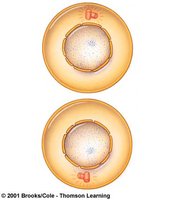
Telophase
Telophase is the final stage of mitosis, where chromosomes decondense and two nuclear membranes form around each set of unduplicated chromosomes.
Chromosome Decondensation: Chromosomes return to their extended form.
Nuclear Envelope Formation: Two nuclei are formed, each with a complete set of chromosomes.
Example: Telophase prepares the cell for cytokinesis and the completion of cell division.
Results of Mitosis
Mitosis results in two daughter nuclei, each with the same chromosome number as the parent cell. Chromosomes are in their unduplicated form, ensuring genetic consistency.
Genetic Consistency: Daughter cells are genetically identical to the parent cell.
Chromosome Number: Maintained across generations of cells.
Example: Mitosis is essential for growth, repair, and maintenance in multicellular organisms.
Additional info:
These notes cover key aspects of cell structure, chromosome organization, and the stages of mitosis, directly relevant to chapters on cell structure and function, cell membrane structure, and cellular reproduction. The included images visually reinforce the concepts of chromosome structure, packing, cell cycle, and mitosis stages.
