 Back
BackCell Signalling and Neuronal Communication: Receptors, Ion Channels, and Action Potentials
Study Guide - Smart Notes
 Tailored notes based on your materials, expanded with key definitions, examples, and context.
Receptors and Cell Signalling
Overview of Cell Signalling
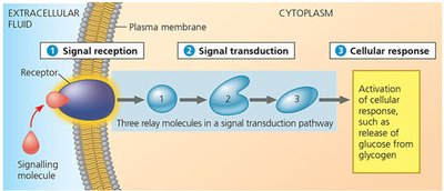
Cell signalling is a fundamental process that allows cells to perceive and respond to their environment. It involves the transmission of signals from the cell surface to the interior, resulting in a specific cellular response. This process is essential for the regulation of cellular activities and coordination among cells in multicellular organisms.
Signal Reception: A signalling molecule (ligand) binds to a specific receptor on the target cell.
Signal Transduction: The receptor activates intracellular relay molecules, transmitting the signal through a cascade of events.
Cellular Response: The cell responds, for example, by altering gene expression, enzyme activity, or cell behavior.
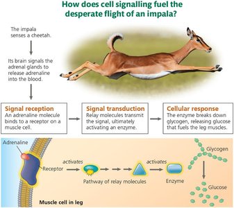
Example: Adrenaline binding to muscle cell receptors triggers glycogen breakdown to fuel muscle contraction during the flight response in animals.
Signalling in Bacteria and Yeast
Unicellular organisms also use chemical signalling to coordinate behavior, especially under environmental stress or during reproduction.
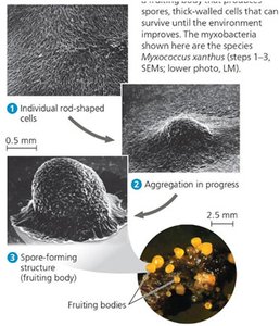
Bacterial Signalling: Myxococcus xanthus bacteria aggregate and form fruiting bodies when nutrients are scarce, using chemical signals to coordinate this process.
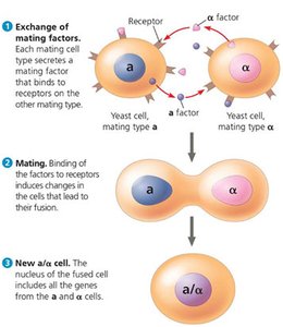
Yeast Signalling: Saccharomyces cerevisiae uses mating factors ('a' and 'α') to identify and fuse with cells of the opposite mating type, leading to genetic recombination.
Types of Cell Signalling
Cell signalling can be classified based on the distance over which the signal acts:
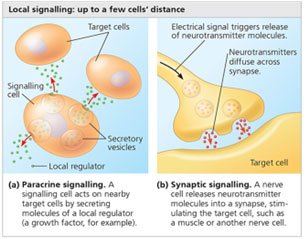
Local Signalling: Includes paracrine (local regulator diffusion) and synaptic signalling (neurotransmitter release at synapses).
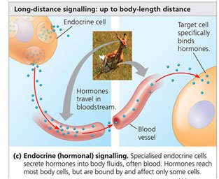
Long-Distance Signalling: Involves hormones released into the bloodstream, affecting distant target cells (endocrine signalling).
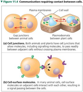
Direct Contact: Cells communicate via gap junctions (animals) or plasmodesmata (plants), or through cell-surface molecules.
Specificity of Signalling
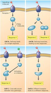
The same signalling molecule can elicit different responses in different cell types due to variations in receptor types and intracellular relay proteins.
Relay proteins and pathways determine the nature of the cellular response.
Cross-talk between pathways and receptor diversity increases signalling complexity.
Receptors, Channels, and Neurons in Cell Signalling
Receptors
Receptors are specialized proteins that detect signalling molecules and initiate cellular responses. They can be located on the cell surface or within the cell.
Ligands: Molecules that bind to receptors, including neurotransmitters, hormones, and drugs.
Types: Extracellular (membrane-bound) and intracellular receptors.
Ligand-Gated Receptors
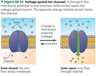
Ligand-gated ion channels open in response to the binding of a specific ligand, allowing ions to flow across the membrane and alter cell function.
Critical for rapid synaptic transmission in neurons.
G-Protein Coupled Receptors (GPCRs)
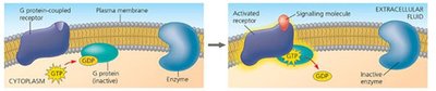
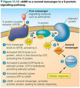
GPCRs are a large family of membrane receptors that activate G-proteins upon ligand binding, triggering intracellular signalling cascades.
Ligand binding causes receptor conformational change, activating the G-protein by exchanging GDP for GTP.
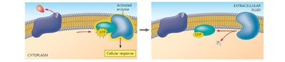
The activated G-protein modulates enzymes or ion channels, leading to a cellular response.
G-protein hydrolyzes GTP to GDP, returning to the inactive state.
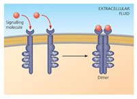
Receptor Tyrosine Kinases (RTKs)
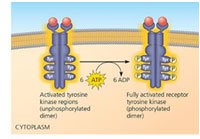
RTKs are membrane receptors that transfer phosphate groups from ATP to tyrosine residues on target proteins, initiating multiple signalling pathways.
Ligand binding induces receptor dimerization and autophosphorylation.
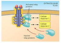
Phosphorylated tyrosines serve as docking sites for relay proteins, triggering diverse cellular responses.
Intracellular Receptors
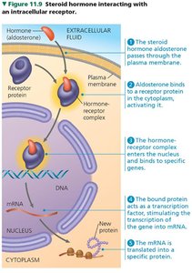
Intracellular receptors are found in the cytoplasm or nucleus and bind small or hydrophobic signalling molecules (e.g., steroid hormones).
The hormone-receptor complex acts as a transcription factor, regulating gene expression.
Ion Channels and Signal Conduction
Major Classes of Ion Channels
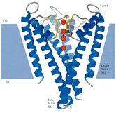
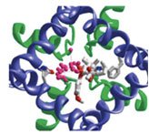
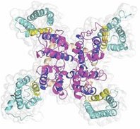
Ion channels are proteins that allow specific ions to pass through the membrane, crucial for generating and propagating electrical signals in neurons.
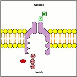
K+ Channels: Voltage-gated and leak channels, important for setting resting membrane potential and repolarization.
Na+ Channels: Open rapidly during depolarization, essential for action potential initiation.
Ca2+ Channels: Involved in neurotransmitter release, muscle contraction, and hormone secretion.
Cl- Channels: Contribute to inhibitory signals and cell volume regulation.
Communication via the Nervous System
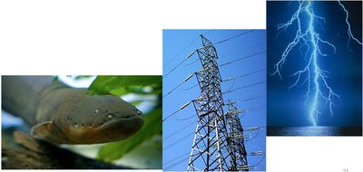
Neurons use electrical signals generated by ion channels to transmit information rapidly and efficiently throughout the body.
Voltage-gated channels change membrane potential, enabling action potentials.
Neuronal communication underlies sensation, movement, and cognition.
Neuron Structure and Types
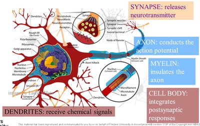
Neurons are specialized cells with distinct regions for receiving, integrating, and transmitting signals.
Dendrites: Receive chemical signals from other neurons.
Cell Body (Soma): Integrates incoming signals.
Axon: Conducts action potentials to target cells.
Synapse: Releases neurotransmitters to communicate with other cells.
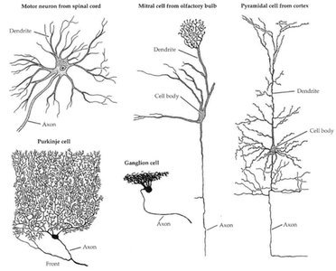
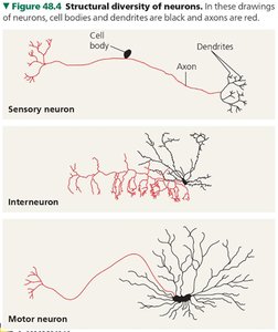
Neurons are classified as sensory, interneurons, or motor neurons based on function.
Ion Movement and Signal Generation
Resting Membrane Potential
The resting membrane potential is the electrical charge difference across the cell membrane, typically ranging from -60 to -90 mV in neurons.
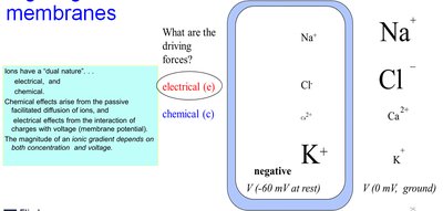
Generated by differential distribution of ions (Na+, K+, Cl-, Ca2+) and selective permeability of the membrane.
Maintained by the Na+/K+-ATPase pump, which moves 3 Na+ out and 2 K+ in per ATP hydrolyzed, creating an electrogenic effect.
Driving forces for ion movement are both electrical (voltage) and chemical (concentration gradient).
Membrane Potential Equation
The membrane potential (Vm) is calculated as:
At rest, Vm typically ranges from -60 to -90 mV.
Glucose Transport and Na+ Gradient
The Na+ gradient established by the Na+/K+-ATPase pump drives the co-transport of glucose into cells. Inhibition of the pump (e.g., by ouabain) decreases glucose uptake over time due to loss of the Na+ gradient.
Action Potentials and Signal Conduction
Action Potential Generation
An action potential is a rapid, transient change in membrane potential that propagates along the axon.
Threshold Potential: The critical level of depolarization required to trigger an action potential.
Depolarization: Opening of voltage-gated Na+ channels causes Na+ influx.
Repolarization: Opening of K+ channels causes K+ efflux.
Hyperpolarization: Membrane potential becomes more negative than resting potential.
Unidirectional Propagation: Due to inactivation of Na+ channels, the action potential moves in one direction.
Equilibrium Potential and Nernst Equation
The equilibrium potential (EX) for an ion X is the membrane voltage at which there is no net flow of that ion. It is calculated using the Nernst equation:
where R is the gas constant, T is temperature, z is the ion charge, F is Faraday's constant, and [X]out and [X]in are the ion concentrations outside and inside the cell, respectively.
Myelination and Saltatory Conduction
Myelin sheaths insulate axons, allowing action potentials to jump between nodes of Ranvier, increasing conduction speed and efficiency (saltatory conduction). Loss of myelin (e.g., in multiple sclerosis) impairs signal transmission.
Action Potential Propagation and Refractory Periods
Action potentials propagate along the axon due to sequential opening and inactivation of voltage-gated Na+ channels. The absolute refractory period ensures unidirectional propagation and limits firing frequency.
Neuronal Communication at Synapses
Synaptic Transmission
When an action potential reaches the end of a neuron (axon terminal), it triggers the release of neurotransmitters into the synaptic cleft. These chemical signals bind to receptors on the postsynaptic cell, opening ligand-gated ion channels and propagating the signal.
Neurotransmitter action is terminated by enzymatic breakdown or reuptake into the presynaptic cell.
Application and Problem Solving
Effects of Channel Blockers
Tetrodotoxin: Blocks voltage-gated Na+ channels, preventing action potential initiation and propagation.
Tetraethylammonium: Blocks voltage-gated K+ channels, prolonging action potential duration by inhibiting repolarization.
Oubain: Inhibits Na+/K+-ATPase, reducing the Na+ gradient and decreasing glucose uptake over time.
Summary Table: Major Receptor Types and Their Mechanisms
Receptor Type | Location | Ligand Example | Mechanism | Cellular Response |
|---|---|---|---|---|
Ligand-Gated Ion Channel | Plasma membrane | Acetylcholine | Opens ion channel upon ligand binding | Rapid change in membrane potential |
G-Protein Coupled Receptor (GPCR) | Plasma membrane | Adrenaline | Activates G-protein, triggers second messenger cascade | Enzyme activation, gene expression changes |
Receptor Tyrosine Kinase (RTK) | Plasma membrane | Growth factors | Dimerization and autophosphorylation | Cell growth, division, differentiation |
Intracellular Receptor | Cytoplasm or nucleus | Steroid hormone | Ligand-receptor complex acts as transcription factor | Gene expression regulation |
