 Back
BackCirculation and Gas Exchange: Structure, Function, and Adaptations
Study Guide - Smart Notes
 Tailored notes based on your materials, expanded with key definitions, examples, and context.
Circulatory Systems and Exchange Surfaces
Diffusion and Simple Body Plans
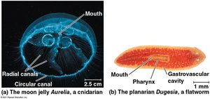
Small molecules move between cells and their surroundings by diffusion, which is efficient only over short distances. Animals with simple body plans, such as cnidarians and flatworms, rely on direct contact between cells and the environment for exchange.
Diffusion: Random thermal motion; time to diffuse increases with the square of the distance.
Simple body plans: Many or all cells are exposed to the environment, minimizing diffusion distance.
Gastrovascular cavity: Functions in both digestion and distribution of substances.
Example: Cnidarians (e.g., Aurelia) and flatworms (Dugesia) have gastrovascular cavities for exchange.
Open vs. Closed Circulatory Systems
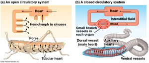
Most animals possess a circulatory system to link exchange surfaces with cells. These systems can be open or closed, each with distinct evolutionary advantages.
Open circulatory system: Circulatory fluid (hemolymph) bathes organs directly; found in insects, arthropods, and some molluscs.
Closed circulatory system: Blood is confined to vessels and is distinct from interstitial fluid; found in annelids, cephalopods, and vertebrates.
Components: Circulatory fluid, interconnecting vessels, muscular pump (heart).
Advantages: Open systems use less energy; closed systems allow larger size, higher activity, and regulated blood distribution.
Organization of Vertebrate Circulatory Systems
Cardiovascular System Structure
Vertebrates have a closed circulatory system called the cardiovascular system, consisting of the heart and blood vessels. Blood flows in one direction through arteries, capillaries, and veins.
Arteries: Carry blood away from the heart; branch into arterioles.
Capillaries: Sites of chemical exchange; form capillary beds.
Veins: Return blood to the heart; converge from venules.
Heart chambers: Blood enters through atria, pumped out through ventricles; chamber number varies among vertebrates.
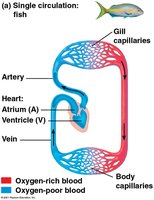
Single Circulation
Single circulation is found in sharks, rays, and bony fishes, characterized by a two-chambered heart. Blood passes through two capillary beds before returning to the heart.
Heart structure: One atrium, one ventricle.
Blood flow: Heart → gill capillaries → body capillaries → heart.
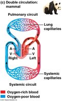
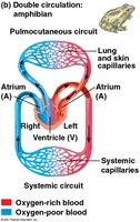
Double Circulation
Double circulation is present in amphibians, reptiles, and mammals. It separates oxygen-poor and oxygen-rich blood, maintaining higher blood pressure in organs.
Pulmonary circuit: Oxygen-poor blood flows to lungs (or lungs and skin in amphibians).
Systemic circuit: Oxygen-rich blood flows to body tissues.
Heart structure: Amphibians: three chambers; mammals and birds: four chambers.
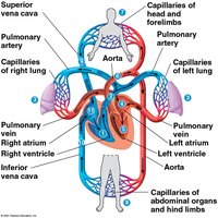
The Mammalian Heart and Cardiac Cycle
Heart Anatomy and Blood Flow
The mammalian heart consists of two atria and two ventricles. The left side handles oxygen-rich blood, while the right side handles oxygen-poor blood.
Blood flow: Right ventricle → pulmonary arteries → lungs → pulmonary veins → left atrium → left ventricle → aorta → body.
Coronary arteries: Supply heart muscle.
Venae cavae: Return oxygen-poor blood to right atrium.
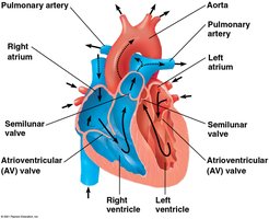
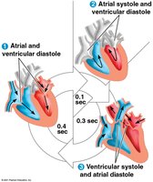
Cardiac Cycle
The heart contracts and relaxes in a rhythmic cycle. Systole is the contraction phase; diastole is the relaxation phase.
Cardiac output: Volume of blood pumped per minute; depends on heart rate and stroke volume.
Valves: AV valves separate atria and ventricles; semilunar valves control flow to aorta and pulmonary artery.
Heart sounds: "Lub-dup" caused by valve closure.
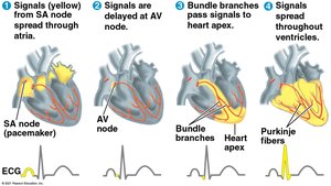
Maintaining Heart Rhythm
Autorhythmic cardiac muscle cells contract without nervous system signals. The sinoatrial (SA) node acts as the pacemaker, setting contraction rate and timing.
Impulse pathway: SA node → AV node (delay) → Purkinje fibers (ventricular contraction).
Regulation: Sympathetic division speeds up, parasympathetic slows down; hormones and temperature also affect rate.
Electrocardiogram (ECG): Records electrical impulses during cardiac cycle.
Blood Vessel Structure and Function
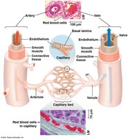
Blood Vessel Anatomy
Blood vessels are structured to match their function. All vessels have a central lumen lined with endothelium.
Capillaries: Thin walls for material exchange.
Arteries: Thick, elastic walls for high pressure.
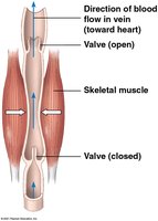
Veins: Thinner walls, valves to prevent backflow.
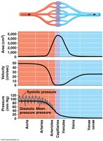
Blood Flow Velocity and Pressure
Blood flow slows in capillaries due to high resistance and large cross-sectional area, then speeds up in veins. Blood pressure is highest in arteries and decreases through the system.
Blood pressure: Force exerted against vessel walls; maintained by elastic recoil of arteries.
Systolic pressure: During ventricular contraction.
Diastolic pressure: During ventricular relaxation.
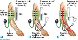
Regulation of Blood Pressure and Gravity
Blood pressure is regulated by altering arteriole diameter (vasoconstriction and vasodilation) and cardiac output. Gravity affects blood pressure, especially in animals with long necks.
Vasoconstriction: Increases blood pressure.
Vasodilation: Decreases blood pressure.
Valves in veins: Prevent backflow; skeletal muscle contraction aids return.
Capillary Function and the Lymphatic System
Capillary Exchange
Exchange of substances occurs across capillary walls. Blood pressure drives fluid out; osmotic pressure pulls fluid in. There is a net loss of fluid from capillaries.
Lymphatic System
The lymphatic system returns leaked fluid (lymph) to the blood, filters it through lymph nodes, and plays a role in defense.
Edema: Swelling from disrupted lymph flow.
Lymph nodes: Filter lymph; swell during infection.
Blood Composition and Function
Blood Components
Blood is a connective tissue with cells suspended in plasma. Plasma contains electrolytes and proteins for osmotic balance, pH buffering, and immunity.
Cellular elements: Erythrocytes (O2 transport), leukocytes (defense), platelets (clotting).
Hemoglobin: Iron-containing protein in erythrocytes; binds O2.
Sickle-cell disease: Abnormal hemoglobin causes sickled cells, leading to blockages and pain.
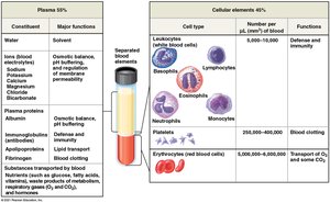
Blood Clotting
Coagulation forms a solid clot from liquid blood. Fibrinogen is converted to fibrin in a cascade of reactions. Thrombus is a clot within a vessel.
Cardiovascular Disease
Atherosclerosis, Heart Attacks, and Stroke
Atherosclerosis is hardening of arteries due to plaque buildup. Heart attacks and strokes result from blocked arteries.
LDL: Delivers cholesterol; high LDL/HDL ratio increases risk.
Statins: Drugs that reduce LDL and heart attack risk.
Hypertension: High blood pressure; controlled by diet, exercise, medication.
Gas Exchange and Respiratory Surfaces
Partial Pressure Gradients
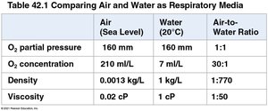
Gas exchange depends on partial pressure gradients. O2 is less soluble in water than air, requiring greater efficiency in aquatic animals.
Property | Air (Sea Level) | Water (20°C) | Air-to-Water Ratio |
|---|---|---|---|
O2 partial pressure | 160 mm | 160 mm | 1:1 |
O2 concentration | 210 ml/L | 7 ml/L | 30:1 |
Density | 0.0013 kg/L | 1 kg/L | 1:770 |
Viscosity | 0.02 cP | 1 cP | 1:50 |
Respiratory Surfaces and Adaptations
Gas exchange occurs by diffusion across respiratory surfaces, which include skin, gills, tracheae, and lungs.
Gills: Outfoldings for aquatic gas exchange; use countercurrent exchange.
Tracheal system: In insects, tubes supply O2 directly to cells.
Lungs: Infoldings for terrestrial gas exchange; rely on circulatory system for transport.
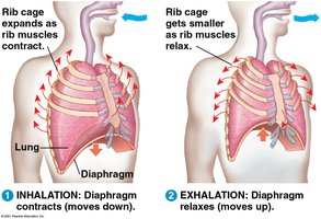
Breathing Mechanisms
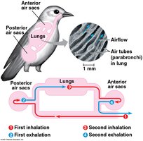
Breathing ventilates the lungs. Amphibians use positive pressure, birds have unidirectional flow with air sacs, and mammals use negative pressure breathing.
Negative pressure breathing: Rib muscles and diaphragm contract to expand lung volume.
Tidal volume: Air inhaled per breath; vital capacity is maximum tidal volume.
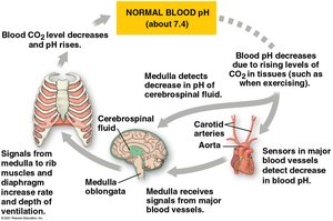
Control of Breathing
Breathing is regulated involuntarily by the medulla oblongata, which responds to pH changes in cerebrospinal fluid and blood.
Sensors: Monitor O2 and CO2 concentrations; signal control centers.
Coordination of Circulation and Gas Exchange
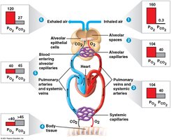
Partial Pressure Gradients in Alveoli
O2 diffuses into blood in alveolar capillaries; CO2 diffuses out. Blood leaving lungs matches alveolar gas pressures.
Respiratory Pigments and Gas Transport
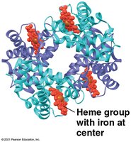
Hemoglobin Structure and Function
Respiratory pigments increase O2 transport capacity. Hemoglobin in vertebrates has four subunits, each with an iron-containing heme group.
Cooperative binding: O2 binding increases affinity in other subunits.
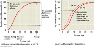
Bohr Shift and CO2 Transport
CO2 lowers blood pH, decreasing hemoglobin's affinity for O2 (Bohr shift). Most CO2 is transported as bicarbonate ions.
Respiratory Adaptations of Diving Mammals
Diving mammals have adaptations for extended underwater periods, including high blood-to-body-volume ratios and myoglobin for O2 storage in muscles. They conserve O2 by passive gliding, selective blood routing, and muscle fermentation.
Example: Weddell seals and Cuvier’s beaked whales demonstrate extreme diving capabilities.
