 Back
BackDNA Structure and Replication: Foundations of Genetic Material
Study Guide - Smart Notes
 Tailored notes based on your materials, expanded with key definitions, examples, and context.
DNA Structure and Replication
Discovery of DNA as Genetic Material
The identification of DNA as the hereditary material was a pivotal moment in biology. Early experiments distinguished DNA from proteins as the carrier of genetic information.
Griffith-Avery Experiment: Demonstrated transformation in bacteria, suggesting a 'transforming principle' (later identified as DNA).
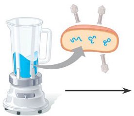
Hershey-Chase Experiment: Used bacteriophages to show that DNA, not protein, enters bacterial cells and directs viral replication.
Watson and Crick: Deduced the double helix structure of DNA, integrating chemical and physical data.
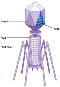
Example: The Hershey-Chase experiment used radioactive isotopes to label DNA and protein, confirming DNA as the genetic material.
DNA Structure
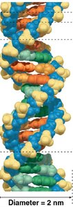
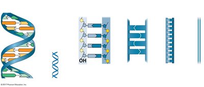
DNA is a double-stranded helical molecule composed of nucleotides. Its structure is essential for its function as genetic material.
Nucleotide: Consists of a phosphate group, deoxyribose sugar, and a nitrogenous base (A, T, G, C).
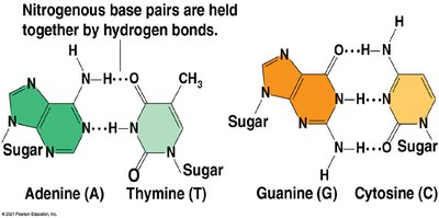
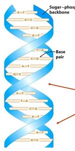
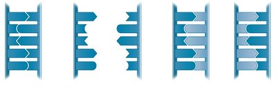
Double Helix: Two antiparallel strands held together by hydrogen bonds between complementary bases.
Base Pairing: Adenine pairs with Thymine (A-T), and Guanine pairs with Cytosine (G-C).
Major and Minor Grooves: The helical structure creates grooves that are important for protein binding.
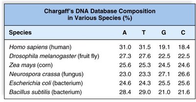
Example: Chargaff's rules state that the amount of A equals T, and G equals C in DNA from any species.
Key Features Defining DNA Structure
Double-stranded helix
Antiparallel orientation (5' to 3' and 3' to 5')
Right-handed helix
Major and minor grooves
DNA Packaging in Chromosomes
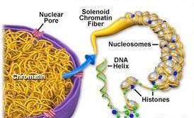
To fit within the nucleus, DNA is highly compacted with proteins into chromatin and chromosomes.
Nucleosome: Basic unit of DNA packaging, consisting of DNA wrapped around histone proteins.
Chromatin: The complex of DNA and proteins that forms chromosomes.
Levels of Compaction: DNA wraps around histones, forming nucleosomes ('beads on a string'), which further coil and fold into higher-order structures.

DNA Replication
Overview of DNA Replication
DNA replication is the process by which a cell duplicates its DNA before cell division. It ensures genetic continuity between generations of cells.
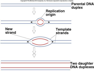
Semiconservative Model: Each new DNA molecule consists of one parental and one newly synthesized strand.
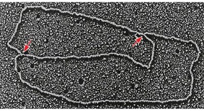
Origin of Replication (ORI): Specific sequence where replication begins.
Replication Fork: The Y-shaped region where the DNA is split into two strands for copying.
Steps of DNA Replication
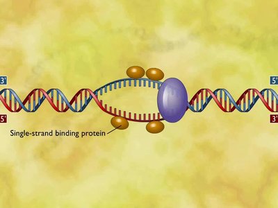
Unwinding the DNA: Helicase separates the two DNA strands at the ORI, creating a replication bubble.
Stabilization: Single-stranded binding proteins (SSBP) prevent re-annealing of the strands.
Relieving Tension: Topoisomerase prevents supercoiling ahead of the replication fork.
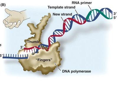
Primer Synthesis: Primase synthesizes a short RNA primer to provide a starting point for DNA polymerase.
DNA Synthesis: DNA polymerase adds nucleotides to the 3' end of the primer, synthesizing the new strand in a 5' to 3' direction.
Leading and Lagging Strands: The leading strand is synthesized continuously, while the lagging strand is synthesized in short Okazaki fragments.
Primer Removal and Ligation: DNA polymerase I removes RNA primers and replaces them with DNA; DNA ligase seals the gaps between fragments.

Leading vs. Lagging Strand Synthesis
Leading Strand: Synthesized continuously toward the replication fork.
Lagging Strand: Synthesized discontinuously away from the fork in Okazaki fragments.
Okazaki Fragments: Short DNA segments on the lagging strand, later joined by DNA ligase.
Limitations and Solutions in DNA Replication
DNA Polymerase Limitations: Cannot initiate synthesis without a primer; can only add nucleotides to the 3' end.
End Replication Problem: Linear chromosomes shorten with each replication due to incomplete synthesis of 5' ends.
Telomeres: Repetitive DNA sequences at chromosome ends that protect coding regions.
Telomerase: Enzyme that extends telomeres in germ cells and some stem cells.
Proofreading and DNA Repair
High fidelity in DNA replication is achieved through proofreading and repair mechanisms.
Proofreading: DNA polymerase checks and corrects errors during synthesis.
Mismatch Repair: Enzymes correct errors missed by DNA polymerase.
Nucleotide Excision Repair: Removes and replaces damaged DNA segments.
Key Terms and Enzymes
Helicase: Unwinds the DNA double helix.
Single-strand binding protein (SSBP): Stabilizes unwound DNA.
Primase: Synthesizes RNA primers.
DNA Polymerase: Synthesizes new DNA strands.
Ligase: Joins Okazaki fragments.
Topoisomerase: Relieves supercoiling.
Telomerase: Extends telomeres.
Summary Table: Leading vs. Lagging Strand
Feature | Leading Strand | Lagging Strand |
|---|---|---|
Synthesis | Continuous | Discontinuous (Okazaki fragments) |
Direction | Toward replication fork | Away from replication fork |
Primer Requirement | One primer | Multiple primers |
DNA Ligase Role | Minimal | Joins fragments |
Additional info:
Mutations arising from replication errors are a source of genetic variation, which is essential for evolution by natural selection.
Shortening of telomeres is associated with cellular aging and cancer biology.
