 Back
BackEngineering the Human Joint: Structure, Function, and Classification
Study Guide - Smart Notes
 Tailored notes based on your materials, expanded with key definitions, examples, and context.
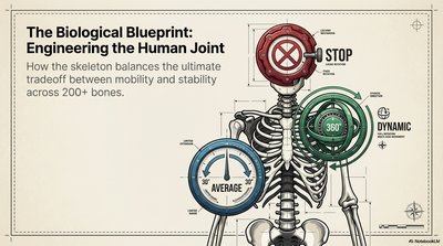
The Biological Blueprint: Engineering the Human Joint
Introduction to Joint Structure and Function
The human skeleton is a marvel of biological engineering, balancing the tradeoff between mobility and stability across more than 200 bones. Joints are the anatomical structures that connect bones, allowing for a range of movements while maintaining structural integrity. Understanding how joints are classified and function is essential for grasping the principles of human movement and skeletal protection.
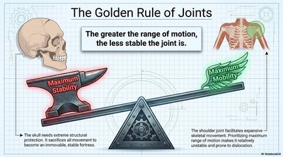
The Golden Rule of Joints
Mobility-Stability Tradeoff
The fundamental principle governing joint design is that the greater the range of motion, the less stable the joint becomes. This tradeoff is evident throughout the skeleton:
Maximum Stability: Joints like those in the skull (sutures) are highly stable but immobile, protecting vital organs such as the brain.
Maximum Mobility: Joints like the shoulder allow for extensive movement but are more prone to instability and dislocation.
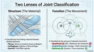
Two Lenses of Joint Classification
Structural and Functional Classification
Joints are classified by two main criteria:
Structure (Material): Based on the binding material between bones.
Fibrous: Dense connective tissue (collagen).
Cartilaginous: Hyaline or fibrocartilage.
Synovial: Fluid-filled capsule.
Function (Movement): Based on the amount of movement allowed.
Synarthrosis (S): Still/stationary, no movement.
Amphiarthrosis (A): Average/little movement.
Diarthrosis (D): Dynamic/free movement.
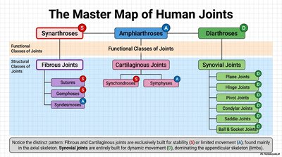
The Master Map of Human Joints
Comprehensive Classification
Human joints are organized into structural and functional classes:
Fibrous Joints | Cartilaginous Joints | Synovial Joints | |
|---|---|---|---|
Synarthroses (S) | Sutures, Gomphoses | Synchondroses | |
Amphiarthroses (A) | Syndesmoses | Symphyses | |
Diarthroses (D) | Plane, Hinge, Pivot, Condylar, Saddle, Ball & Socket |
Pattern: Fibrous and cartilaginous joints are built for stability or limited movement, while synovial joints are specialized for dynamic movement, especially in the limbs.
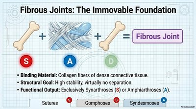
Fibrous Joints: The Immovable Foundation
Structure and Function
Binding Material: Collagen fibers of dense connective tissue.
Structural Goal: High stability, virtually no separation between bones.
Functional Output: Exclusively synarthroses (immovable) or amphiarthroses (slightly movable).
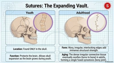
Sutures: The Expanding Vault
Location: Found only in the skull.
Function: Protects the brain and allows skull expansion during youth.
Form: Wavy, interlocking edges add strength; ossify with age to form a bony joint (synostosis).
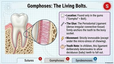
Gomphoses: The Living Bolts
Location: Found only in the gums (teeth sockets).
Glue: Periodontal ligament anchors tooth to bone.
Movement: Strictly immovable except for micro-stress during chewing.
Youth Note: Ligament deteriorates in children to allow baby teeth to fall out.
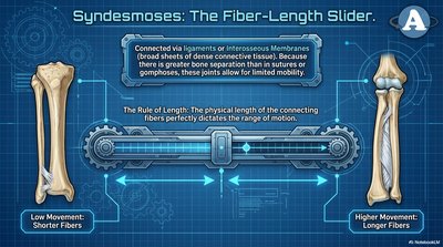
Syndesmoses: The Fiber-Length Slider
Connection: Ligaments or interosseous membranes (dense connective tissue).
Movement: Limited mobility; range depends on fiber length (longer fibers = more movement).
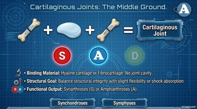
Cartilaginous Joints: The Middle Ground
Structure and Function
Binding Material: Hyaline cartilage or fibrocartilage; no joint cavity.
Structural Goal: Balance between structural integrity and slight flexibility or shock absorption.
Functional Output: Synarthroses or amphiarthroses.
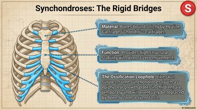
Synchondroses: The Rigid Bridges
Material: Bones bound by hyaline cartilage.
Function: High stability, almost zero movement.
Ossification Loophole: Some synchondroses (e.g., growth plates) are temporary and replaced by bone.
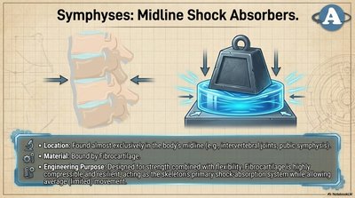
Symphyses: Midline Shock Absorbers
Location: Found in the body's midline (e.g., intervertebral joints, pubic symphysis).
Material: Bound by fibrocartilage.
Purpose: Strength with flexibility; fibrocartilage acts as a shock absorber while allowing limited movement.
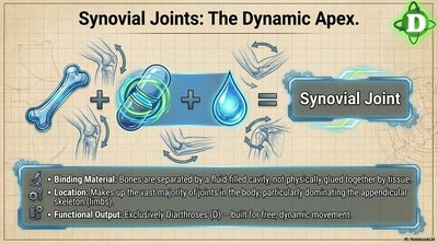
Synovial Joints: The Dynamic Apex
Structure and Function
Binding Material: Bones separated by a fluid-filled cavity, not glued by tissue.
Location: Most joints in the body, especially in the limbs.
Functional Output: Exclusively diarthroses (free movement).
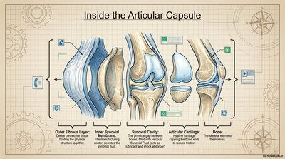
Inside the Articular Capsule
Outer Fibrous Layer: Dense connective tissue, adds strength and stability.
Inner Synovial Membrane: Produces synovial fluid for lubrication and shock absorption.
Synovial Cavity: Space filled with synovial fluid.
Articular Cartilage: Covers bone surfaces, reduces friction.
Bone: Skeletal elements forming the joint.
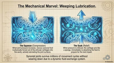
The Mechanical Marvel: Weeping Lubrication
The Squeeze (Compression): Synovial fluid is forced out of cartilage into the cavity, lubricating the joint.
The Soak (Relief): Cartilage reabsorbs fluid when pressure is relieved, maintaining joint health.
The Blueprint Synthesis: Form Meets Function
Summary Table of Joint Types
Synarthroses (Red S) | Amphiarthroses (Blue A) | Diarthroses (Green D) | |
|---|---|---|---|
Fibrous | Sutures (Skull), Gomphoses (Teeth) | Syndesmoses (Interosseous membranes) | |
Cartilaginous | Synchondroses (Costosternal) | Symphyses (Spine/Midline) | |
Synovial | All Synovial Joints (Knees, Shoulders, Limbs) |
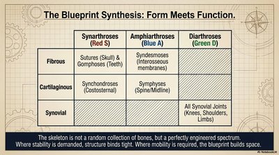
Conclusion: The skeleton is a spectrum of engineered joints, where stability is achieved through strong binding structures and mobility is enabled by specialized joint spaces. This organization allows the human body to perform a vast array of movements while protecting vital organs and maintaining structural integrity.
