 Back
BackImmunology: The Lymphatic System and Immune Defenses
Study Guide - Smart Notes
 Tailored notes based on your materials, expanded with key definitions, examples, and context.
The Lymphatic System
Structure and Function of the Lymphatic System
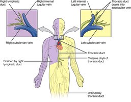
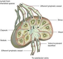
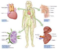
The lymphatic system is a network of vessels and organs that plays a crucial role in immune defense and fluid balance. It collects excess tissue fluid (lymph), absorbs fats from the small intestine, and returns lymph to the bloodstream via the subclavian veins.
Lymphatic capillaries absorb excess tissue fluid and fats.
Lymphatic veins transport lymph to ducts that drain into the subclavian veins.
Lymph nodes filter lymph and store lymphocytes and other white blood cells.
Defense Against Infections
Primary (Innate) Defense Mechanisms
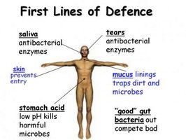
Primary defenses are non-specific barriers that prevent pathogens from entering the body. These include physical, chemical, and biological barriers.
Skin: Acts as a physical barrier to pathogens.
Oil glands: Secrete chemicals that deter bacterial growth.
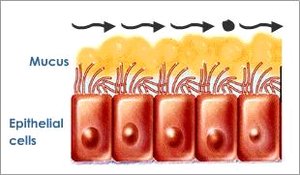
Mucous membranes and cilia: Trap and remove pathogens from passages exposed to the environment.
Acidic pH: Certain body regions (e.g., stomach, skin) have low pH to kill pathogens.
Indigenous bacteria: Compete with pathogens for resources and may release inhibitory chemicals.
Saliva and tears: Contain lysozymes that digest microbial cell walls.

Secondary (Innate) Defense: The Inflammatory Response
Mechanism of Inflammation
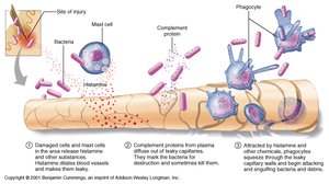
The inflammatory response is activated when pathogens breach primary defenses. It involves a series of events that localize infection and recruit immune cells to the site of injury.
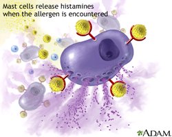
Histamines released by damaged cells and mast cells dilate capillaries, increasing blood and lymph flow.
Protein complements (chemokines) attract white blood cells (phagocytes) to the infection site.
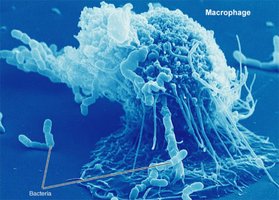
Phagocytes engulf microbes and dead cells, cleaning and disinfecting tissue.
Bradykinins stimulate nerve endings, causing pain.
Signs of inflammation: redness, swelling, heat, and pain.

Specific (Adaptive) Immunity
Lymphocytes: B Cells and T Cells
Specific immunity involves lymphocytes that recognize and respond to specific antigens. B cells and T cells are the main types of lymphocytes, each with distinct roles.
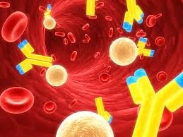
B lymphocytes (B cells): Mature in bone marrow; secrete antibodies into blood and lymph (humoral immunity).
T lymphocytes (T cells): Mature in thymus; differentiate into helper T cells and cytotoxic T cells (cell-mediated immunity).
Lymphocyte Activation
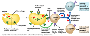
Lymphocyte activation is initiated when a macrophage presents a foreign antigen to a helper T cell, which then coordinates the immune response.
Macrophages present a "self-non-self" complex on their surface after engulfing a pathogen.
Helper T cells recognize this complex and are activated.
Activated helper T cells secrete cytokines to stimulate other immune cells:
Interleukin 4: Activates B cells for humoral response.
Interleukin 12: Activates cytotoxic T cells for cell-mediated immunity.
Cell-Mediated Immunity
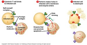
Cytotoxic T cells destroy infected cells by releasing toxins such as perforin, which creates holes in the target cell membrane, leading to apoptosis (programmed cell death).
Activated cytotoxic T cells bind to infected cells displaying foreign antigens.
Perforin forms pores in the infected cell membrane.
Enzymes enter and induce apoptosis.
Humoral (Acquired) Immunity
Humoral immunity is mediated by antibodies produced by B cells. It is activated by exposure to antigens and is responsible for defending against pathogens in body fluids.
Antigens trigger antibody production and the inflammatory response.
Acquired immunity "remembers" antigens for faster future responses (basis of vaccination).
Vaccines introduce harmless antigens to stimulate immunity without causing disease.
Antigens and Antibodies
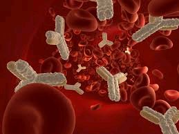
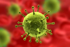
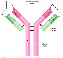
Antigens are molecules (usually proteins or polysaccharides) on the surface of pathogens that are recognized by the immune system. Antibodies are Y-shaped proteins that specifically bind to antigens, neutralizing them and marking them for destruction.
Antibodies have two antigen-binding sites and a heavy chain tail.
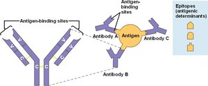
They recognize specific regions on antigens called antigenic determinants or epitopes.
Antigen-Antibody Complex and Elimination Mechanisms
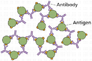
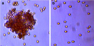
The formation of antigen-antibody complexes neutralizes pathogens and marks them for elimination by phagocytes. Antibodies can also cause agglutination (clumping) of pathogens and activate complement proteins to lyse foreign cells.
Neutralization: Antibodies block pathogen activity.
Agglutination: Antibodies cause pathogens to clump, facilitating phagocytosis.
Complement activation: Leads to lysis of foreign cells.
Additional info: These notes cover the structure and function of the lymphatic system, innate and adaptive immune defenses, and the mechanisms by which the body recognizes and eliminates pathogens. They are suitable for exam preparation in a college-level biology course, specifically for chapters on the immune system and defense mechanisms.
