 Back
BackMembrane Structure and Function: A Comprehensive Study Guide
Study Guide - Smart Notes
 Tailored notes based on your materials, expanded with key definitions, examples, and context.
Membrane Structure and Function
Overview: Life at the Edge
The plasma membrane is a fundamental cellular structure that acts as a selective barrier, regulating the movement of substances into and out of the cell. Its selective permeability is essential for maintaining homeostasis and supporting cellular life.
Cellular Membranes: Fluid Mosaics of Lipids and Proteins
Phospholipid Structure and Amphipathic Nature
Phospholipids are the most abundant lipids in the plasma membrane. They are amphipathic molecules, meaning they contain both hydrophobic (water-fearing) tails and hydrophilic (water-loving) heads. This dual nature allows them to form bilayers that are the foundation of all cellular membranes.
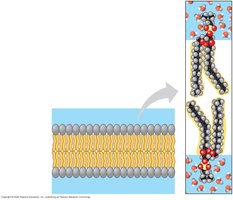
Hydrophilic heads face outward toward aqueous environments.
Hydrophobic tails face inward, shielded from water.
Fluid Mosaic Model: This model describes the membrane as a fluid structure with a mosaic of various proteins embedded within or attached to the bilayer.
The Fluidity of Membranes
Phospholipids and some proteins can move laterally within the bilayer, contributing to membrane fluidity. Rarely, phospholipids may flip-flop transversely across the membrane.
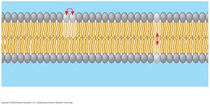
Lateral movement: Occurs frequently, allowing dynamic rearrangement.
Flip-flop: Rare, involves movement from one leaflet to the other.
Factors Affecting Membrane Fluidity
Membrane fluidity is influenced by temperature, the composition of fatty acids, and the presence of cholesterol.
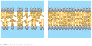
Decreasing temperature reduces fluidity.
Unsaturated hydrocarbon tails (with kinks) increase fluidity.
Saturated hydrocarbon tails make the membrane more viscous.
Cholesterol acts as a fluidity buffer:
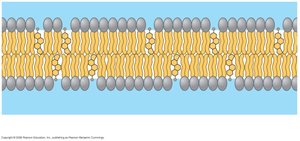
At high temperatures, cholesterol restrains movement of phospholipids.
At low temperatures, it prevents tight packing, thus preventing solidification.
Membrane Proteins and Their Functions
Types of Membrane Proteins
Membrane proteins are embedded in or associated with the lipid bilayer and are responsible for most of the membrane's specific functions.
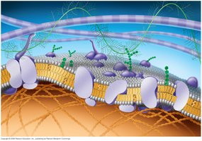
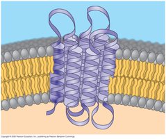
Integral proteins: Penetrate the hydrophobic core, often spanning the membrane.
Peripheral proteins: Loosely bound to the membrane surface.
Glycoproteins and glycolipids: Involved in cell recognition and signaling.
Major Functions of Membrane Proteins
Membrane proteins perform six major functions:
Transport: Move substances across the membrane.
Enzymatic activity: Catalyze specific reactions.
Signal transduction: Relay signals from outside to inside the cell.
Cell-cell recognition: Identify and interact with other cells.
Intercellular joining: Connect adjacent cells.
Attachment to the cytoskeleton and ECM: Maintain cell shape and stabilize protein location.
Membrane Structure and Selective Permeability
The Permeability of the Lipid Bilayer
The plasma membrane is selectively permeable, allowing some substances to cross more easily than others.
Hydrophobic (nonpolar) molecules (e.g., hydrocarbons) pass through rapidly.
Polar molecules (e.g., sugars) and ions do not cross easily without assistance.
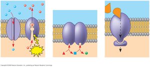
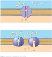
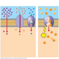
Transport Proteins
Transport proteins facilitate the movement of hydrophilic substances across the membrane. There are two main types:
Channel proteins: Provide hydrophilic tunnels for specific molecules or ions (e.g., aquaporins for water).
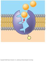
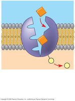
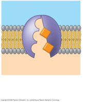
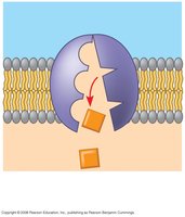
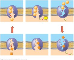
Carrier proteins: Bind to molecules and change shape to shuttle them across the membrane.
Passive Transport: Diffusion and Facilitated Diffusion
Diffusion
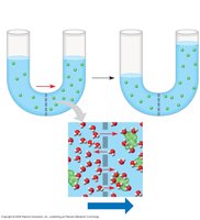
Diffusion is the movement of molecules from an area of higher concentration to an area of lower concentration, down their concentration gradient. It is a passive process that does not require energy input.
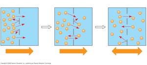
At dynamic equilibrium, molecules continue to move, but there is no net change in concentration.
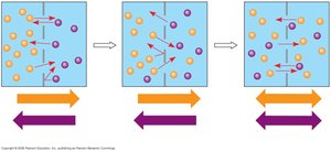
Osmosis
Osmosis is the diffusion of water across a selectively permeable membrane. Water moves from regions of lower solute concentration to regions of higher solute concentration.
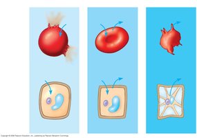
Water Balance of Cells Without Walls
Tonicity describes the ability of a surrounding solution to cause a cell to gain or lose water:
Isotonic: No net water movement; cell remains stable.
Hypertonic: Cell loses water; shrivels.
Hypotonic: Cell gains water; may lyse (burst).
Osmoregulation
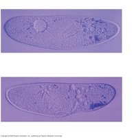
Osmoregulation is the control of water balance, crucial for organisms in hypertonic or hypotonic environments. For example, the protist Paramecium uses a contractile vacuole to expel excess water.
Facilitated Diffusion
Facilitated diffusion is passive transport aided by proteins. Channel proteins provide corridors for specific molecules or ions, while carrier proteins undergo shape changes to move substances across the membrane.
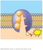
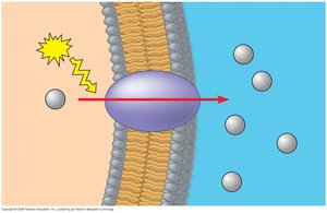
Active Transport: Moving Against the Gradient
Sodium-Potassium Pump
Active transport uses energy (usually ATP) to move solutes against their concentration gradients. The sodium-potassium pump is a classic example, exchanging Na+ and K+ ions across the plasma membrane.
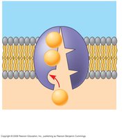
For each cycle, 3 Na+ ions are pumped out and 2 K+ ions are pumped in.
This process helps maintain the cell's electrochemical gradient.
Electrochemical Gradient and Electrogenic Pumps
The electrochemical gradient is the combined effect of the ion's concentration gradient and the membrane potential. Electrogenic pumps, such as the sodium-potassium pump in animals and the proton pump in plants, fungi, and bacteria, generate voltage across membranes.
Types of Membrane Transport
Type | Energy Required? | Direction | Example |
|---|---|---|---|
Simple Diffusion | No | Down gradient | O2, CO2 |
Facilitated Diffusion | No | Down gradient | Glucose, ions |
Active Transport | Yes (ATP) | Against gradient | Na+/K+ pump |
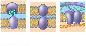
Bulk Transport: Exocytosis and Endocytosis
Exocytosis
Exocytosis is the process by which cells export large molecules by fusing vesicles with the plasma membrane, releasing their contents outside the cell.
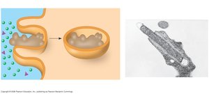
Endocytosis
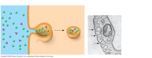
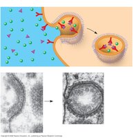
Endocytosis is the process by which cells import large molecules by engulfing them in vesicles formed from the plasma membrane. There are three main types:
Phagocytosis: "Cellular eating"; cell engulfs large particles or cells.
Pinocytosis: "Cellular drinking"; cell takes in extracellular fluid and dissolved solutes.
Receptor-mediated endocytosis: Specific molecules are taken in after binding to receptors.
Additional info: This guide expands on the lecture outline by providing definitions, examples, and a summary table for types of membrane transport, ensuring a comprehensive and self-contained resource for exam preparation.
