 Back
BackBio 100 LEC Chapter 7 Part 2
Study Guide - Smart Notes
 Tailored notes based on your materials, expanded with key definitions, examples, and context.
Bio 100 LEC Chapter 7 Part 2
Membrane Structure and Function
Overview
This chapter explores the structure and function of biological membranes, focusing on their selective permeability, transport mechanisms, and the physiological consequences of membrane transport. The plasma membrane is essential for regulating the exchange of materials between the cell and its environment, maintaining cellular homeostasis, and supporting cell function.
Membrane Structure
Phospholipid Bilayer and Fluidity
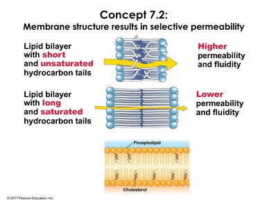
The plasma membrane is primarily composed of a phospholipid bilayer, which provides both structural integrity and selective permeability. The properties of phospholipids, such as the length and saturation of hydrocarbon tails, directly affect membrane fluidity and permeability.
Short, unsaturated hydrocarbon tails increase membrane fluidity and permeability.
Long, saturated hydrocarbon tails decrease membrane fluidity and permeability.
Cholesterol modulates membrane fluidity by preventing tight packing of phospholipids.
Selective Permeability of the Lipid Bilayer
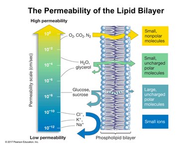
Membrane permeability depends on the size and polarity of molecules:
Small, nonpolar molecules (e.g., O2, CO2, N2) diffuse easily through the bilayer.
Small, uncharged polar molecules (e.g., H2O, glycerol) have moderate permeability.
Large, uncharged polar molecules (e.g., glucose, sucrose) are largely restricted.
Ions (e.g., Na+, K+, Cl-) cannot cross without assistance due to their charge and hydration shells.
Transport Proteins
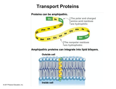
Transport proteins facilitate the movement of substances across the membrane. They are amphipathic, containing both hydrophilic and hydrophobic regions, allowing integration into the lipid bilayer.
Hydrophilic regions interact with polar heads and aqueous environments.
Hydrophobic regions interact with the hydrophobic core of the bilayer.
Transmembrane proteins span the entire bilayer and are essential for transport.
Aquaporins
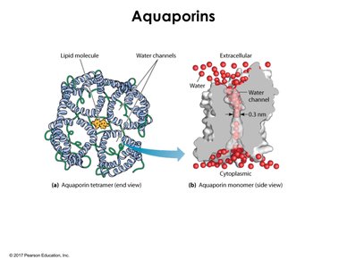
Aquaporins are specialized channel proteins that facilitate rapid movement of water across the membrane. They are tetrameric transmembrane proteins, with a polar channel allowing water to pass single file.
Channel size: ~0.3 nm, restricting passage to water molecules only.
Polar amino acids line the channel, supporting water transport.
Function: Increases water permeability compared to simple diffusion.
Passive Transport
Simple Diffusion
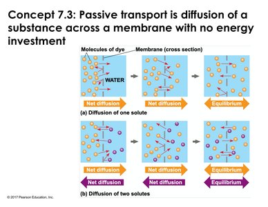
Simple diffusion is the movement of molecules from high to low concentration across the membrane without energy input. The process continues until equilibrium is reached.
Concentration gradient: Drives diffusion.
Equilibrium: Achieved when solute concentrations are equal on both sides.
Osmosis
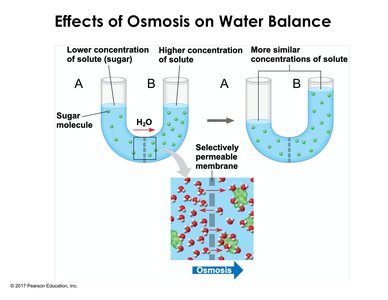
Osmosis is the diffusion of water across a selectively permeable membrane. Water moves from areas of lower solute concentration to higher solute concentration, adjusting volumes to reach equilibrium.
Selective permeability: Allows water but not solute (e.g., sugar) to cross.
Result: Water moves to equalize solute concentrations.
Osmolarity and Water Balance in Cells
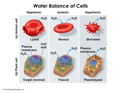
Osmolarity refers to the total solute concentration. The relationship between cell and environment determines water movement:
Hypotonic solution: Lower solute outside; water enters cell. Animal cells may lyse, plant cells become turgid.
Isotonic solution: Equal solute; no net water movement. Animal cells remain normal, plant cells become flaccid.
Hypertonic solution: Higher solute outside; water leaves cell. Animal cells shrivel, plant cells undergo plasmolysis.
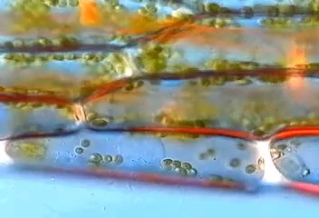
Microscopic Effects of Osmosis in Plant Cells
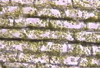
Microscopic images illustrate the effects of hypertonic and hypotonic solutions on plant cells:
Hypertonic: Cytoplasm pulls away from cell wall, chloroplasts cluster centrally (plasmolysis).
Hypotonic: Cells become firm and turgid, chloroplasts expand, cell wall remains unchanged.
Facilitated Diffusion
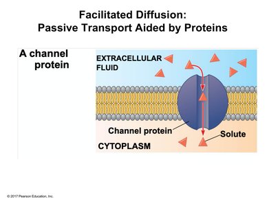
Channel Proteins
Channel proteins provide corridors for specific molecules or ions to move down their concentration gradient. Examples include aquaporins and ion channels.
Open/closed states: Channels may require stimuli to open.
Specificity: Channels are selective for certain solutes.
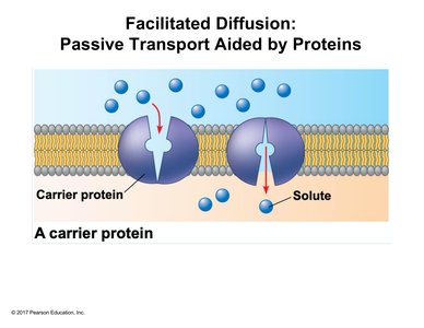
Carrier Proteins
Carrier proteins facilitate diffusion by undergoing conformational changes upon solute binding, allowing translocation across the membrane.
Shape change: Triggered by solute binding and release.
Direction: Always down the concentration gradient.
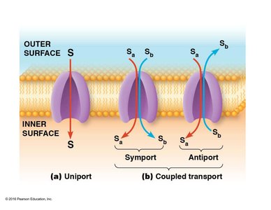
Types of Carrier Protein Transport
Carrier proteins can transport one or more solutes:
Uniport: Single solute, one direction.
Symport: Two solutes, same direction.
Antiport: Two solutes, opposite directions.
Active Transport
Overview of Active Transport
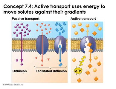
Active transport moves solutes against their concentration gradients, requiring energy input (usually ATP). Carrier proteins are essential for this process.
Energy source: ATP hydrolysis.
Direction: Against concentration gradient.
Carrier proteins: Required for active transport.
Sodium-Potassium Pump
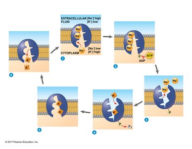
The sodium-potassium pump is a classic example of active transport, maintaining cellular ion gradients and membrane potential.
Steps:
Three Na+ ions bind to the pump facing the cytoplasm.
ATP hydrolysis phosphorylates the pump.
Conformational change releases Na+ outside.
Two K+ ions bind from outside.
Phosphate group is released, pump returns to cytoplasmic orientation, K+ released inside.
Electrogenic: Generates voltage across the membrane.
Membrane Potential and Electrochemical Gradients
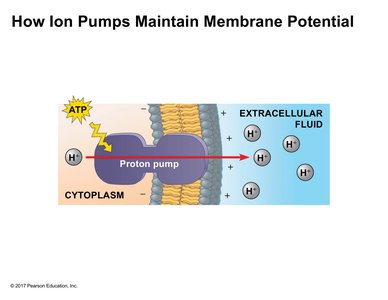
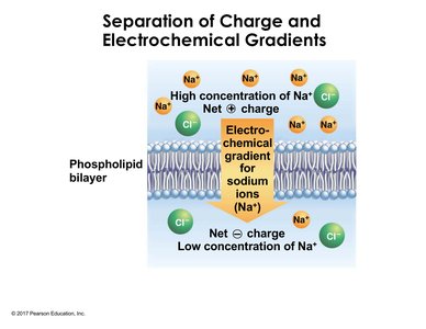
Membrane potential is the voltage across the membrane, established by ion pumps. Electrochemical gradients combine electrical and concentration gradients to drive ion movement.
Membrane potential: Difference in charge across membrane.
Electrochemical gradient: Combination of membrane potential and ion concentration.
Secondary Active Transport (Cotransport)
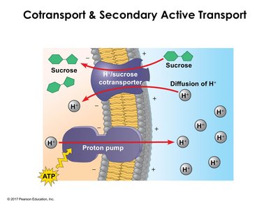
Secondary active transport uses the energy from one solute moving down its gradient to drive another solute against its gradient. Example: H+/sucrose cotransporter.
Proton pump: Uses ATP to create H+ gradient.
Cotransporter: Couples H+ influx with sucrose import.
Indirect ATP use: ATP is used by the pump, not the cotransporter.
Bulk Transport: Endocytosis and Exocytosis
Overview of Bulk Transport
Bulk transport moves large or polar substances across the membrane via vesicle formation. Endocytosis brings substances into the cell, while exocytosis expels them.
Phagocytosis: Cell eating; engulfment of particles.
Pinocytosis: Cell drinking; uptake of extracellular fluid.
Receptor-mediated endocytosis: Specific uptake via membrane receptors.
Receptor-Mediated Endocytosis and Disease
Receptor-mediated endocytosis is crucial for the uptake of specific molecules, such as LDL cholesterol. Genetic defects in LDL receptor function can lead to hypercholesterolemia and increased risk of heart disease.
Normal cells: Efficient LDL uptake and breakdown.
Mutant cells: Reduced or absent LDL uptake, leading to high circulating LDL.
Summary Table: Types of Membrane Transport
Transport Type | Energy Required | Direction | Protein Involved | Example |
|---|---|---|---|---|
Simple Diffusion | No | Down gradient | No | O2 diffusion |
Facilitated Diffusion | No | Down gradient | Yes (channel/carrier) | Aquaporin, ion channels |
Active Transport | Yes (ATP) | Against gradient | Yes (carrier) | Sodium-potassium pump |
Bulk Transport | Yes | Variable | Yes (vesicle formation) | Endocytosis, exocytosis |
Key Equations
Diffusion: Fick's Law of Diffusion Where J is flux, D is diffusion coefficient, dC/dx is concentration gradient.
Osmosis: Osmotic Pressure Where \Pi is osmotic pressure, i is van't Hoff factor, M is molarity, R is gas constant, T is temperature.
