 Back
BackMembrane Structure and Function: Chapter 7 Study Guide
Study Guide - Smart Notes
 Tailored notes based on your materials, expanded with key definitions, examples, and context.
Membrane Structure and Function
Overview
This chapter explores the structure and function of cellular membranes, emphasizing their role in compartmentalization, regulation, and communication within and between cells. The plasma membrane is a dynamic, selectively permeable barrier composed of lipids, proteins, and carbohydrates, crucial for maintaining cellular integrity and facilitating essential biological processes.

The Functions of Membranes
Main Roles of Biological Membranes
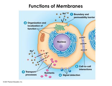
Biological membranes are essential for cellular function, providing boundaries, regulating permeability, and supporting communication and organization within cells.
Boundary and Permeability Barrier: Membranes define cell edges and regulate the movement of substances, allowing selective entry and exit.
Organization and Localization of Function: Membranes partition cellular regions, enabling specialized environments for distinct biochemical processes (e.g., mitochondria, Golgi apparatus).
Transport Processes: Membranes house proteins that facilitate the movement of ions and nutrients, such as the sodium-potassium pump and sugar transporters.
Signal Detection: Membrane proteins detect external signals and initiate intracellular responses, forming the basis of cell communication.
Cell-to-Cell Interactions: Membranes enable direct communication and solute exchange between adjacent cells, vital for tissues like plant cells and cardiac muscle.
Membrane Regulation of Cellular Traffic
Mechanisms of Transport
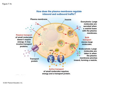
The plasma membrane regulates the movement of molecules through various mechanisms, including passive and active transport, and bulk transport.
Passive Transport: Movement of molecules down their concentration gradient without energy input; may involve transport proteins.
Active Transport: Movement against concentration gradients, requiring energy (usually ATP) and transport proteins.
Bulk Transport: Movement of large molecules via vesicles, including exocytosis (secretion) and endocytosis (uptake).
Membrane Structure: Fluid Mosaic Model
Components and Organization
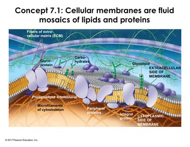
Cellular membranes are fluid mosaics composed of lipids, proteins, and carbohydrates. The arrangement and diversity of these molecules contribute to membrane function and asymmetry.
Phospholipids: Amphipathic molecules forming the bilayer, with hydrophilic heads and hydrophobic tails.
Cholesterol: Modulates membrane fluidity and stability.
Proteins: Integral and peripheral proteins serve roles in transport, signaling, and structural support.
Carbohydrates: Attached to lipids (glycolipids) or proteins (glycoproteins), important for cell recognition and signaling.
Asymmetry: Different molecules are distributed between the inner and outer layers, affecting function and communication.
Synthesis and Sidedness of Membranes
Membrane Assembly and Modification
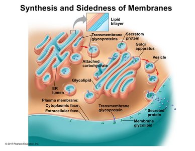
Membranes are synthesized and modified in the endoplasmic reticulum (ER) and Golgi apparatus, establishing distinct inside and outside faces. Glycoproteins and glycolipids are sorted and transported via vesicles to their destinations.
ER: Initial synthesis and glycosylation of proteins and lipids.
Golgi Apparatus: Further modification and sorting for membrane integration or secretion.
Vesicle Transport: Movement of membrane components to the plasma membrane, maintaining sidedness and functional specificity.
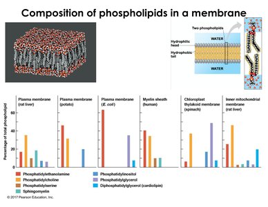
Phospholipid Diversity and Distribution
Types and Functions of Phospholipids
Phospholipids vary in head groups and tail composition, contributing to membrane diversity across cell types and organisms. Their amphipathic nature is fundamental to bilayer formation and membrane function.
Head Groups: Examples include choline, inositol, and sphingosine (e.g., sphingomyelin).
Distribution: Varies between plasma membrane, mitochondrial membrane, and other organelles.
Amphipathic Properties: Hydrophilic heads interact with water; hydrophobic tails form the membrane core.
Membrane Fluidity
Movement of Lipids and Proteins
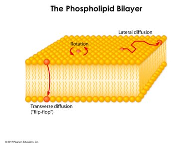
Membranes are dynamic, allowing lateral movement of lipids and proteins. Fluidity is essential for membrane function, including transport and signaling.
Lateral Diffusion: Lipids and proteins move within the same layer.
Rotation: Individual molecules rotate in place.
Transverse Diffusion (Flip-Flop): Rare movement between layers.
Experimental Evidence for Fluidity
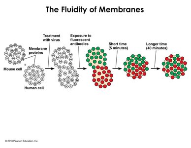
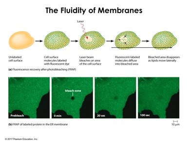
Experiments such as cell fusion and FRAP (fluorescent recovery after photobleaching) demonstrate the dynamic nature of membranes.
Cell Fusion: Mixing of membrane proteins from different cells shows lateral mobility.
FRAP: Recovery of fluorescence in bleached areas confirms lateral diffusion of proteins.
Factors Affecting Membrane Fluidity
Saturation, Cholesterol, and Tail Length
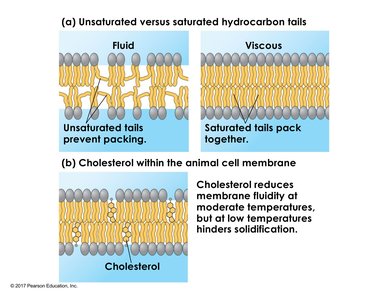
Membrane fluidity is influenced by the saturation of fatty acid tails, cholesterol content, and tail length.
Unsaturated Tails: Cis double bonds create kinks, increasing fluidity.
Saturated Tails: Allow tight packing, increasing viscosity and reducing fluidity.
Cholesterol: Acts as a fluidity buffer, decreasing fluidity at high temperatures and preventing solidification at low temperatures.
Tail Length: Longer tails increase hydrophobic interactions, reducing fluidity.
Homeoviscous Adaptation
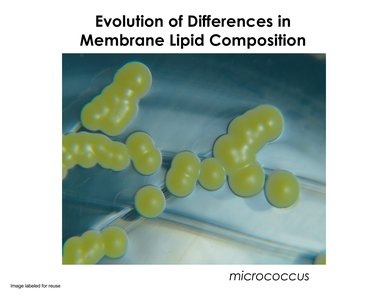
Organisms regulate membrane fluidity by altering lipid composition, especially in response to temperature changes. For example, bacteria like Micrococcus activate enzymes to shorten fatty acid tails at lower temperatures, maintaining fluidity.
Membrane Proteins: Types and Functions
Functional Roles
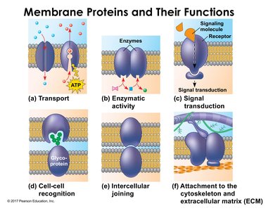
Membrane proteins are responsible for transport, enzymatic activity, signal transduction, cell recognition, intercellular joining, and attachment to the cytoskeleton and extracellular matrix.
Transport: Move substances across membranes, often using ATP.
Enzymatic Activity: Catalyze reactions at the membrane surface.
Signal Transduction: Relay external signals to internal cellular responses.
Cell-Cell Recognition: Glycoproteins enable identification and interaction between cells.
Intercellular Joining: Proteins link cells together for tissue formation.
Attachment: Connect membrane to cytoskeleton and extracellular matrix for structural support.
Medical Example: HIV Entry
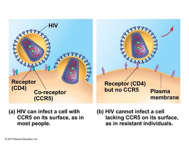
HIV entry into host cells depends on membrane proteins (CD4 and CCR5 receptors). Individuals lacking CCR5 are resistant to HIV infection, illustrating the critical role of membrane proteins in health and disease.
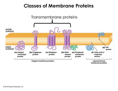
Classes of Membrane Proteins
Membrane proteins are classified based on their association with the bilayer:
Integral Proteins: Penetrate the hydrophobic core; include monotopic and transmembrane proteins.
Peripheral Proteins: Attach to membrane surfaces via non-covalent interactions.
Lipid-Anchored Proteins: Covalently attached to lipids, anchoring them in the membrane.
Selective Permeability of Membranes
Concept and Factors
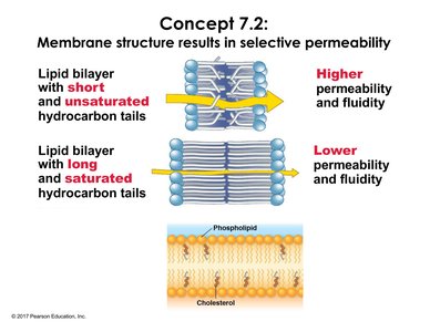
Membrane structure results in selective permeability, allowing certain molecules to pass while restricting others. Fluidity and permeability are closely linked, influenced by lipid composition.
Short, Unsaturated Tails: Increase permeability and fluidity.
Long, Saturated Tails: Decrease permeability and fluidity.
Permeability of the Lipid Bilayer
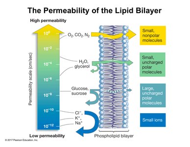
The lipid bilayer is most permeable to small, nonpolar molecules (e.g., O2, CO2, N2), less permeable to small uncharged polar molecules (e.g., H2O, glycerol), and largely impermeable to large uncharged polar molecules and ions without assistance.
Small Nonpolar Molecules: Highest permeability.
Small Uncharged Polar Molecules: Moderate permeability.
Large Uncharged Polar Molecules: Low permeability.
Small Ions: Very low permeability; require transport proteins.
Summary Table: Membrane Functions and Properties
Function | Key Components | Example |
|---|---|---|
Boundary & Permeability | Phospholipids, proteins | Regulation of Na+ entry |
Organization | Membrane-bound organelles | Mitochondria, Golgi |
Transport | Transport proteins | Sodium-potassium pump |
Signal Detection | Receptor proteins | Hormone signaling |
Cell-Cell Interaction | Junction proteins | Gap junctions in heart |
