 Back
BackMembrane Structure and Function: Fluidity, Permeability, and Transport
Study Guide - Smart Notes
 Tailored notes based on your materials, expanded with key definitions, examples, and context.
Membrane Structure and Function
Fluid Mosaic Model of Cellular Membranes
The plasma membrane is a dynamic, complex structure essential for cellular function. It is primarily composed of lipids, proteins, and carbohydrates, organized according to the fluid mosaic model.
Phospholipids: Amphipathic molecules with hydrophilic heads and hydrophobic tails, forming a stable bilayer.
Membrane Proteins: Amphipathic, with hydrophilic regions exposed to water and hydrophobic regions embedded in the bilayer. Includes integral (transmembrane) and peripheral proteins.
Carbohydrates: Attached to proteins (glycoproteins) or lipids (glycolipids), serving as cell recognition markers.
Cholesterol: Acts as a fluidity buffer, stabilizing membrane structure across temperature changes.
Fluid Mosaic Model: Describes the membrane as a mosaic of proteins floating in a fluid phospholipid bilayer.
Example: The experiment by Frye and Edidin demonstrated lateral movement of membrane proteins by fusing mouse and human cells and observing marker mixing.
Membrane Fluidity and Evolutionary Adaptations
Membrane fluidity is crucial for permeability and protein function, influenced by lipid composition and environmental conditions.
Unsaturated Hydrocarbon Tails: Enhance fluidity at lower temperatures due to kinks from double bonds.
Saturated Hydrocarbon Tails: Increase viscosity and solidify at higher temperatures.
Cholesterol: Reduces fluidity at moderate temperatures, prevents solidification at low temperatures.
Evolutionary Adaptations: Organisms adjust membrane lipid composition to maintain fluidity (e.g., fishes in cold environments have more unsaturated lipids).
Membrane Proteins: Types and Functions
Membrane proteins are responsible for most membrane functions and are classified as integral or peripheral.
Integral Proteins: Penetrate the hydrophobic interior, often spanning the membrane (transmembrane).
Peripheral Proteins: Loosely bound to the membrane surface.
Functions:
Transport: Channels and carriers move substances across the membrane.
Enzymatic Activity: Catalyze reactions at the membrane surface.
Signal Transduction: Relay messages from external signals.
Cell-Cell Recognition: Glycoproteins serve as identification tags.
Intercellular Joining: Form junctions between cells.
Attachment: Connect to cytoskeleton and ECM for structural support.
Example: HIV resistance is linked to the absence of the CCR5 co-receptor, preventing viral entry.
Membrane Carbohydrates and Cell-Cell Recognition
Carbohydrates on the extracellular surface of the plasma membrane are crucial for cell recognition and distinction.
Glycoproteins and Glycolipids: Short, branched carbohydrate chains attached to proteins or lipids.
Diversity: Carbohydrate composition varies among species, individuals, and cell types.
Example: Human blood types (A, B, AB, O) are determined by glycoprotein carbohydrate variations.
Synthesis and Sidedness of Membranes
Membranes have distinct inside and outside faces, established during synthesis and transport through the ER and Golgi apparatus.
Step 1: Proteins and lipids synthesized in the ER; carbohydrates added to proteins.
Step 2: Further modification in the Golgi; formation of glycolipids.
Step 3: Transported in vesicles to the plasma membrane.
Step 4: Vesicle fusion (exocytosis) positions carbohydrates on the extracellular face.
Membrane Permeability and Transport
Selective Permeability of Membranes
Biological membranes allow some substances to cross more easily than others, regulating transport and maintaining cellular homeostasis.
Nonpolar Molecules: (e.g., hydrocarbons, O2, CO2) can dissolve in the lipid bilayer and cross easily.
Polar Molecules and Ions: (e.g., glucose, water, ions) require transport proteins due to the hydrophobic interior.
Transport Proteins: Channel and carrier proteins facilitate movement of specific substances.
Transport Proteins: Channel and Carrier Types
Transport proteins enable selective movement of ions and polar molecules across membranes.
Channel Proteins: Provide hydrophilic tunnels (e.g., aquaporins for water).
Carrier Proteins: Bind and change shape to shuttle molecules across.
Specificity: Each transport protein is selective for particular substances.
Passive Transport: Diffusion and Osmosis
Diffusion and Dynamic Equilibrium
Diffusion is the passive movement of molecules from high to low concentration, resulting in dynamic equilibrium.
Concentration Gradient: Drives diffusion without energy input.
Dynamic Equilibrium: Equal movement in both directions, no net change.
Osmosis and Tonicity
Osmosis is the diffusion of water across a selectively permeable membrane, influenced by solute concentration and tonicity.
Tonicity: Effect of solute concentration on water movement.
Isotonic: Equal solute concentration; no net water movement.
Hypotonic: Lower solute concentration outside; water enters cell.
Hypertonic: Higher solute concentration outside; water leaves cell.
Water Potential (Ψ): Indicates the tendency of water to move; measured in bars or megapascals.
Low solute concentration = high water potential.
High solute concentration = low water potential.
Water moves from high to low water potential.
Osmolarity: Total solute concentration per liter; high osmolarity = low water potential.
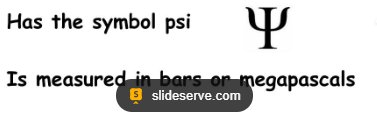
Example: Water moves from areas of high water potential (low solute) to low water potential (high solute).
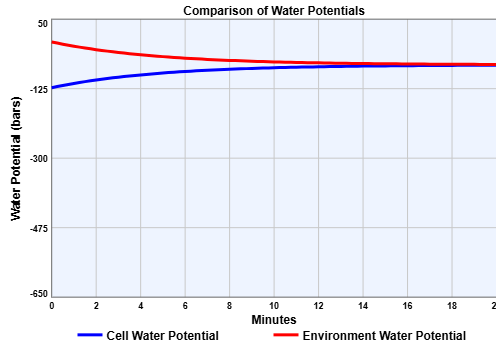
Example: The graph shows water potential changes in a cell and its environment, illustrating dynamic equilibrium as potentials converge.
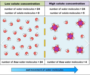
Example: Diagram shows that free water molecules move across the membrane, while bound water molecules (attached to solutes) cannot.
Facilitated Diffusion
Facilitated diffusion is passive transport aided by proteins, allowing polar molecules and ions to cross membranes efficiently.
Channel Proteins: Aquaporins and ion channels provide rapid passage.
Carrier Proteins: Undergo shape changes to move solutes down their gradient.
Gated Channels: Open or close in response to stimuli, crucial in nerve cells.
Active Transport and Bulk Transport
Active Transport: Moving Solutes Against Gradients
Active transport uses energy (usually ATP) to move solutes against their concentration gradients, maintaining internal cellular conditions.
Carrier Proteins: Essential for active transport.
Sodium-Potassium Pump: Exchanges Na+ and K+ across the membrane, powered by ATP.
Electrogenic Pumps: Generate voltage across membranes (e.g., Na/K pump in animals, proton pump in plants).
Cotransport: Coupled transport of two solutes; one moves down its gradient, driving the uphill movement of another.
Example: Glucose and Na+ cotransport in intestinal cells enables efficient nutrient absorption.
Bulk Transport: Exocytosis and Endocytosis
Large molecules are transported across membranes in bulk via vesicles, through exocytosis and endocytosis.
Exocytosis: Vesicles fuse with the plasma membrane, releasing contents outside the cell.
Endocytosis: Cell takes in molecules by forming vesicles from the plasma membrane.
Phagocytosis: Engulfs particles (cellular eating).
Pinocytosis: Takes in fluid (cellular drinking).
Receptor-mediated Endocytosis: Specific uptake of substances via receptor proteins.
Membrane Rejuvenation: Exocytosis and endocytosis remodel and maintain plasma membrane integrity.
Example: LDL uptake by receptor-mediated endocytosis; defects cause familial hypercholesterolemia.
Summary Table: Tonicity and Water Potential
Condition | Solute Concentration | Water Movement | Cell Effect |
|---|---|---|---|
Isotonic | Equal inside and outside | No net movement | Stable cell volume |
Hypotonic | Lower outside | Water enters cell | Cell swells/lyses (animal); turgid (plant) |
Hypertonic | Higher outside | Water leaves cell | Cell shrivels (animal); plasmolysis (plant) |
Key Equations
Water Potential Equation:
Where is total water potential, is solute potential, and is pressure potential.
Osmolarity:
Additional info: Water potential is a central concept in plant physiology, determining the direction of water movement and influencing processes such as transpiration and nutrient uptake.
