 Back
BackBio 100 LEC Chapter 7 Part 1
Study Guide - Smart Notes
 Tailored notes based on your materials, expanded with key definitions, examples, and context.
Bio 100 LEC Chapter 7 Part 1
Membrane Structure and Function
Introduction to Membrane Structure and Function
The plasma membrane is a fundamental component of all cells, serving as a dynamic boundary that regulates the internal environment and mediates communication with the external environment. Understanding membrane structure and function is essential for grasping cellular processes, including transport, signaling, and compartmentalization.
Major Functions of Biological Membranes
Compartmentalization and Communication
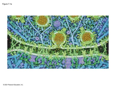
Biological membranes are not merely static barriers; they are dynamic structures that facilitate compartmentalization, regulate molecular traffic, and enable cell-to-cell communication. Membranes create distinct environments within cells, allowing specialized functions to occur efficiently and safely.
Compartmentalization: Membranes form boundaries that separate cellular regions, supporting the localization of specific biochemical processes.
Regulation: The plasma membrane selectively permits the movement of substances, maintaining homeostasis and preventing inappropriate cellular responses.
Communication: Membranes enable the fusion of vesicles and the transfer of signaling molecules between cells, crucial for processes such as neurotransmission.
Key Functions of Membranes
Membranes perform several essential roles in cellular physiology, as summarized below:
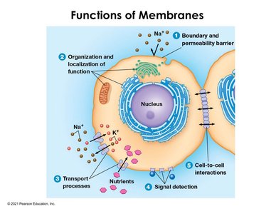
Boundary and Permeability Barrier: Defines the cell's limits and regulates entry and exit of substances.
Organization and Localization of Function: Segregates cellular processes into organelles (e.g., mitochondria, Golgi apparatus).
Transport Processes: Facilitates the movement of ions and nutrients via transport proteins (e.g., sodium-potassium pump).
Signal Detection: Membrane proteins detect external signals and initiate intracellular responses (signal transduction).
Cell-to-Cell Interactions: Enables direct communication and material exchange between adjacent cells (e.g., gap junctions in animal cells, plasmodesmata in plants).
Membrane Regulation of Traffic
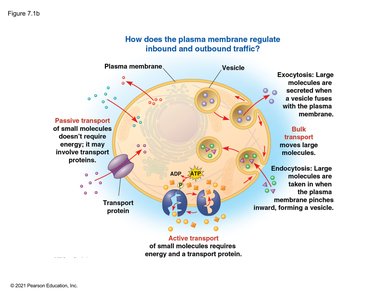
The plasma membrane regulates the movement of molecules through various mechanisms, including passive and active transport, as well as bulk transport processes such as endocytosis and exocytosis.
Passive Transport: Movement of molecules down their concentration gradient without energy input (e.g., diffusion, facilitated diffusion).
Active Transport: Movement of molecules against their concentration gradient, requiring energy (usually ATP) and transport proteins.
Bulk Transport: Large molecules are transported via vesicles in processes such as endocytosis (import) and exocytosis (export).
Membrane Structure: The Fluid Mosaic Model
Fluid Mosaic Model
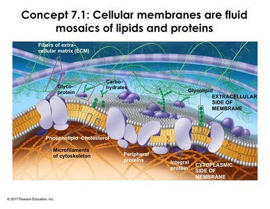
Cellular membranes are described by the fluid mosaic model, which highlights their composition of lipids, proteins, and carbohydrates. The membrane is a dynamic, two-dimensional fluid where components can move laterally.
Lipids: Primarily phospholipids, which are amphipathic molecules with hydrophilic heads and hydrophobic tails.
Proteins: Integral and peripheral proteins are embedded or associated with the lipid bilayer, performing various functions.
Carbohydrates: Attached to lipids (glycolipids) or proteins (glycoproteins), mainly on the extracellular surface, involved in cell recognition and signaling.
Asymmetry: The two leaflets of the bilayer differ in lipid and protein composition, reflecting functional specialization.
Synthesis and Sidedness of Membranes
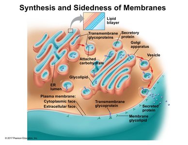
Membrane asymmetry is established during synthesis in the endoplasmic reticulum (ER) and Golgi apparatus. Proteins and lipids are modified and sorted, resulting in distinct inner and outer membrane faces.
ER and Golgi: Synthesize and modify membrane proteins and lipids, adding carbohydrate groups to form glycoproteins and glycolipids.
Vesicular Transport: Vesicles shuttle membrane components to their destinations, preserving sidedness.
Phospholipids: Structure and Diversity
Phospholipid Structure and Types
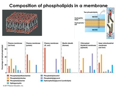
Phospholipids are the most abundant membrane lipids, characterized by two nonpolar fatty acid tails and a polar head group. Variations in head groups and backbone (glycerol or sphingosine) contribute to membrane diversity.
Amphipathic Nature: Drives the spontaneous formation of bilayers in aqueous environments.
Examples: Phosphatidylcholine, phosphatidylethanolamine, sphingomyelin (animal cells), and others.
Distribution: Phospholipid composition varies between organisms, cell types, and organelles, reflecting functional requirements.
Phospholipid Bilayer Dynamics
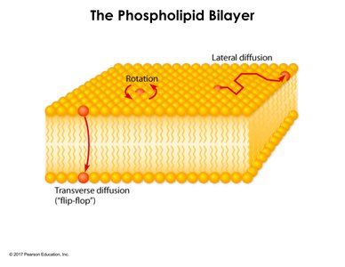
The phospholipid bilayer is held together by weak hydrophobic interactions, allowing for significant lateral movement of lipids and some proteins. Types of movement include:
Lateral Diffusion: Lipids move side-to-side within the same leaflet.
Rotation: Individual phospholipids rotate in place.
Transverse Diffusion (Flip-Flop): Rare movement of lipids between leaflets due to energetic barriers.
Experimental Evidence for Membrane Fluidity
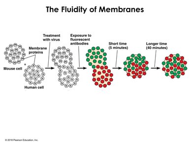
Membrane fluidity has been demonstrated experimentally using cell fusion and fluorescence techniques:
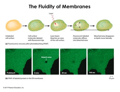
Cell Fusion Experiments: Fusion of cells from different species, labeled with fluorescent antibodies, shows mixing of membrane proteins over time, supporting lateral mobility.
FRAP (Fluorescence Recovery After Photobleaching): A region of membrane is bleached with a laser, and recovery of fluorescence indicates lateral diffusion of proteins and lipids.
Factors Affecting Membrane Fluidity
Fatty Acid Composition and Cholesterol
Membrane fluidity is influenced by the types of fatty acids and the presence of cholesterol:
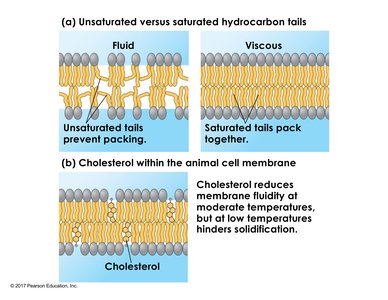
Unsaturated Fatty Acids: Contain cis double bonds that introduce kinks, preventing tight packing and increasing fluidity.
Saturated Fatty Acids: Lack double bonds, allowing tight packing and decreasing fluidity (increasing viscosity).
Trans Fats: Behave similarly to saturated fats, increasing membrane viscosity.
Cholesterol: Acts as a fluidity buffer, reducing fluidity at high temperatures and preventing solidification at low temperatures by disrupting regular packing of phospholipids.
Fatty Acid Tail Length: Longer tails increase hydrophobic interactions, reducing fluidity.
Homeoviscous Adaptation
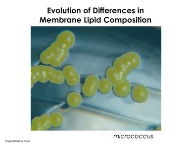
Organisms can adjust membrane lipid composition to maintain optimal fluidity under changing temperatures, a process known as homeoviscous adaptation. For example, the bacterium Micrococcus shortens fatty acid tails at lower temperatures to preserve membrane fluidity.
Membrane Proteins: Structure and Function
Functions of Membrane Proteins
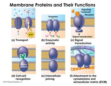
Membrane proteins are responsible for most of the specific functions of membranes. Major categories include:
Transport: Move substances across the membrane (e.g., channels, pumps).
Enzymatic Activity: Catalyze reactions at the membrane surface.
Signal Transduction: Relay signals from the external environment to the cell interior.
Cell-Cell Recognition: Glycoproteins serve as identification tags for cellular interactions.
Intercellular Joining: Proteins link adjacent cells together (e.g., tight junctions, gap junctions).
Attachment: Anchor the membrane to the cytoskeleton and extracellular matrix, maintaining cell shape and stability.
Medical Application: HIV Entry and Membrane Proteins
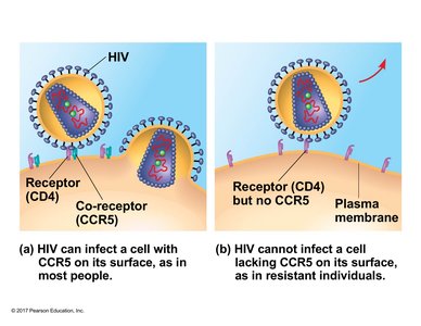
The importance of membrane proteins is illustrated by the entry of HIV into host cells. HIV requires binding to the CD4 receptor and a co-receptor (CCR5) on the host cell membrane. Individuals lacking functional CCR5 are resistant to HIV infection, demonstrating the critical role of membrane proteins in disease susceptibility.
Classes of Membrane Proteins
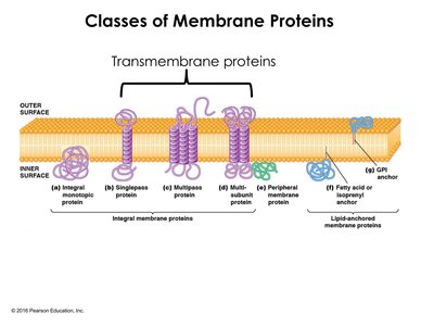
Membrane proteins are classified based on their association with the lipid bilayer:
Integral Membrane Proteins: Penetrate the hydrophobic core; include monotopic (one side) and transmembrane (span the bilayer) proteins. Transmembrane proteins may cross the membrane once (single-pass) or multiple times (multi-pass).
Peripheral Membrane Proteins: Loosely bound to the membrane surface or to integral proteins via non-covalent interactions; do not penetrate the hydrophobic core.
Lipid-Anchored Proteins: Covalently attached to lipids that insert into the membrane, anchoring the protein.
Integral and lipid-anchored proteins require disruption of the membrane for removal, while peripheral proteins can be dissociated more easily.
Additional info: This guide integrates foundational concepts from Chapter 7 of a standard biology textbook, emphasizing the structure, composition, and functional diversity of cellular membranes. It also connects to related topics such as cell communication, organelle function, and experimental techniques in membrane biology.
