 Back
BackMembrane Transport and Cell Signaling: Structure, Function, and Mechanisms
Study Guide - Smart Notes
 Tailored notes based on your materials, expanded with key definitions, examples, and context.
Membrane Structure and Function
Overview: Life at the Edge
The plasma membrane is a fundamental cellular structure that separates the internal environment of the cell from its external surroundings. It is selectively permeable, allowing certain substances to cross more easily than others, thus maintaining homeostasis.
Plasma membrane: Defines cell boundaries and regulates molecular traffic.
Selectively permeable: Permits passage of some molecules while restricting others.
Fluid Mosaic Model
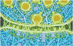
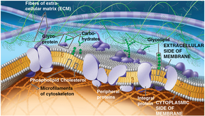
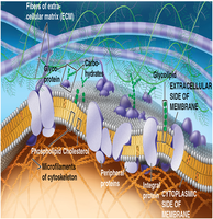
The plasma membrane is primarily composed of amphipathic lipids (phospholipids), proteins, and carbohydrates. The fluid mosaic model describes the membrane as a dynamic structure with proteins floating in or on a fluid lipid bilayer.
Amphipathic lipids: Have both hydrophobic (water-fearing) and hydrophilic (water-loving) regions.
Proteins: Integral and peripheral proteins serve various functions, including transport and signaling.
Carbohydrates: Attached to proteins (glycoproteins) or lipids (glycolipids), mainly on the extracellular surface.
Phospholipid Bilayer Structure
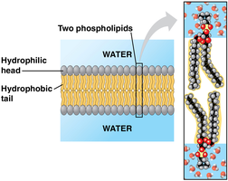
The basic structure of the membrane is a bilayer of phospholipids, with hydrophilic heads facing outward toward water and hydrophobic tails facing inward, away from water. This arrangement forms a semi-permeable barrier.
Hydrophilic heads: Interact with aqueous environments inside and outside the cell.
Hydrophobic tails: Face each other, forming the membrane's core.
Membrane Fluidity
Lipid and Protein Movement
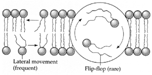
Membrane lipids and some proteins are not fixed in place; they move laterally within the layer, contributing to membrane fluidity. Rarely, lipids may flip-flop between layers.
Lateral movement: Frequent and rapid movement of lipids within the same layer.
Flip-flop: Rare movement of lipids from one layer to the other.
Factors Affecting Fluidity
Temperature: Higher temperatures increase fluidity; lower temperatures decrease it.
Lipid composition: Unsaturated fatty acids increase fluidity due to kinks in their tails; saturated fatty acids decrease fluidity by packing tightly.
Cholesterol: Acts as a fluidity buffer, reducing fluidity at high temperatures and preventing tight packing at low temperatures.
Membrane Proteins and Their Functions
Types of Membrane Proteins
Membrane proteins are essential for various cellular functions and are classified based on their association with the lipid bilayer.
Integral proteins: Penetrate the hydrophobic core; many are transmembrane proteins with both hydrophobic and hydrophilic regions.
Peripheral proteins: Loosely bound to the membrane surface, not embedded in the lipid bilayer.
Functions of Membrane Proteins
Transport: Move substances across the membrane.
Enzymatic activity: Catalyze chemical reactions.
Signal transduction: Relay signals from outside to inside the cell.
Cell-cell recognition: Identify and interact with other cells.
Intercellular joining: Connect adjacent cells.
Attachment to cytoskeleton and ECM: Maintain cell shape and stabilize membrane proteins.
Membrane Carbohydrates and Cell Recognition
Glycolipids and Glycoproteins
Carbohydrates attached to lipids (glycolipids) or proteins (glycoproteins) are found on the extracellular surface of the plasma membrane. They play a key role in cell-cell recognition and communication.
Glycolipids: Carbohydrates covalently bonded to lipids.
Glycoproteins: Carbohydrates covalently bonded to proteins.
Membrane Permeability and Transport
Selective Permeability
The lipid bilayer is selectively permeable, allowing hydrophobic (nonpolar) molecules to cross easily, while hydrophilic (polar) molecules require assistance from transport proteins.
Easily cross: O2, CO2 (hydrophobic, small molecules)
Require assistance: Glucose, water (large, polar, or charged molecules)
Transport Proteins
Channel proteins: Provide hydrophilic tunnels for specific molecules or ions (e.g., aquaporins for water).
Carrier proteins: Bind to molecules and change shape to shuttle them across the membrane (e.g., glucose carrier protein).
Passive Transport
Diffusion
Diffusion is the movement of molecules from an area of high concentration to an area of low concentration, driven by the concentration gradient. Passive transport requires no energy input.
Dynamic equilibrium: Achieved when net movement ceases, but molecules continue to move randomly.
Osmosis
Osmosis is the diffusion of water across a selectively permeable membrane, moving from lower solute concentration to higher solute concentration until equilibrium is reached.
Tonicity and Water Balance
Hypotonic solution: Lower solute concentration outside; cell gains water and may burst (lysis).
Isotonic solution: Equal solute concentration; no net water movement.
Hypertonic solution: Higher solute concentration outside; cell loses water and shrivels.
Plant Cells and Water Balance
Turgid: Plant cell in hypotonic solution; cell swells but is protected by cell wall.
Flaccid: Plant cell in isotonic solution; cell becomes limp.
Plasmolysis: Plant cell in hypertonic solution; membrane pulls away from cell wall, usually lethal.
Facilitated Diffusion
Facilitated diffusion is passive transport aided by proteins. Channel and carrier proteins help specific molecules move down their concentration gradients without energy input.
Channel proteins: Provide corridors for ions or molecules (e.g., ion channels, aquaporins).
Carrier proteins: Undergo shape changes to transport molecules (e.g., glucose transporters).
Active Transport
Mechanism and Importance
Active transport moves substances against their concentration gradients, from low to high concentration, using energy (usually ATP). This process maintains essential differences in ion concentrations across membranes.
Sodium-potassium pump: Exchanges Na+ for K+ across animal cell membranes, maintaining membrane potential.
Membrane Potential and Ion Pumps
Membrane potential: Voltage across a membrane due to unequal distribution of ions.
Electrochemical gradient: Combination of electrical and chemical forces driving ion movement.
Electrogenic pump: Transport protein that generates voltage (e.g., sodium-potassium pump in animals, proton pump in plants, fungi, and bacteria).
Cotransport
Cotransport occurs when the downhill diffusion of one solute drives the uphill transport of another. For example, in plants, the proton gradient established by proton pumps is used to drive the active transport of nutrients.
Bulk Transport
Exocytosis and Endocytosis
Large molecules cross the membrane in bulk via vesicles, a process requiring energy.
Exocytosis: Vesicles fuse with the plasma membrane to release contents outside the cell.
Endocytosis: Cell takes in molecules by forming vesicles from the plasma membrane. Types include:
Phagocytosis: "Cellular eating" of large particles.
Pinocytosis: "Cellular drinking" of extracellular fluid.
Receptor-mediated endocytosis: Specific uptake of molecules via receptor proteins.
Cell Signaling
Local and Long-Distance Signaling
Cells communicate to coordinate activities, often involving the plasma membrane. Signaling can be local (e.g., paracrine, synaptic) or long-distance (e.g., endocrine/hormonal).
Direct contact: Gap junctions (animals) and plasmodesmata (plants) allow cytosolic exchange.
Paracrine signaling: Local signaling via secreted molecules.
Synaptic signaling: Nerve cells release neurotransmitters to target cells.
Endocrine signaling: Hormones travel through the circulatory system to distant cells.
Stages of Cell Signaling
Reception: Detection of signaling molecule by receptor protein.
Transduction: Signal is relayed and amplified through a cascade of molecular interactions.
Response: Cellular activity is altered, such as gene expression or enzyme activity.
Membrane Receptors
G protein-coupled receptors: Transmit signals via G proteins.
Ligand-gated ion channels: Open or close in response to ligand binding, allowing ion flow.
Signal Transduction Pathways
Signal transduction often involves multiple steps, amplifying the signal and allowing for regulation and coordination of cellular responses. The final response may involve changes in gene expression or cytoplasmic activity.
Summary Table: Types of Membrane Transport
Transport Type | Energy Required? | Direction | Example |
|---|---|---|---|
Simple Diffusion | No | Down gradient | O2, CO2 |
Facilitated Diffusion | No | Down gradient | Glucose, water (via aquaporins) |
Active Transport | Yes (ATP) | Against gradient | Sodium-potassium pump |
Bulk Transport | Yes | In or out | Exocytosis, endocytosis |
