 Back
BackNeurons, Synapses, and Signaling: Structure and Function of Nervous Systems
Study Guide - Smart Notes
 Tailored notes based on your materials, expanded with key definitions, examples, and context.
Neurons, Synapses, and Signaling
Introduction to Nervous Systems
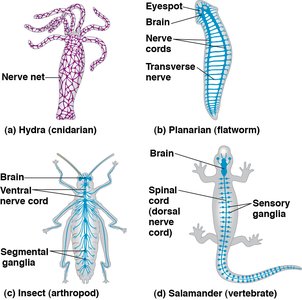
The nervous system is a complex network responsible for processing information and coordinating responses in animals. Nervous systems range from simple nerve nets in cnidarians to highly centralized brains in vertebrates, enabling advanced behaviors such as learning and memory.
Cephalization: The evolutionary concentration of nervous tissue and sensory organs at the anterior end (head) of the body, facilitating forward movement and predation.
Ganglia: Clusters of nerve cell bodies that process sensory information and coordinate motor responses in simpler animals.
Brain: A centralized organ capable of integrating complex sensory inputs and generating sophisticated behaviors.
Neuron Structure and Organization
Form and Function of Neurons
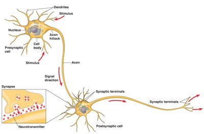
Neurons are specialized cells that exemplify the relationship between structure and function. They are organized to receive, process, and transmit information efficiently.
Cell Body (Soma): Contains the nucleus and metabolic machinery.
Dendrites: Highly branched extensions that receive signals from other neurons.
Axon: A long extension that transmits signals to other cells, ending in axon terminals.
Synapse: The junction where an axon terminal communicates with another cell, separated by a synaptic cleft.
Neurotransmitters: Chemical messengers released from vesicles in the axon terminal to transmit signals across the synapse.
Glial Cells: Neuron Support
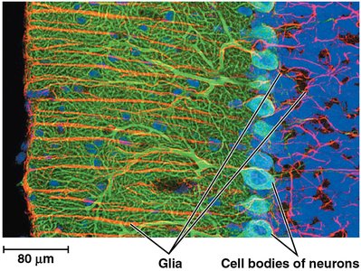
Glial cells, or glia, are non-neuronal cells that support, nourish, and protect neurons. In the mammalian brain, glia outnumber neurons by 10- to 50-fold.
Functions: Nourishment, insulation (myelin sheath), regulation of extracellular fluid, and guidance during development.
Types: Astrocytes (blood-brain barrier), oligodendrocytes (CNS myelin), Schwann cells (PNS myelin).
Types of Neurons and Information Processing
Functional Classes of Neurons
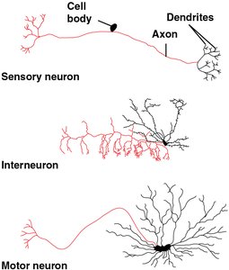
Animal nervous systems are composed of three main types of neurons, each with distinct roles in processing information:
Sensory Neurons: Detect external or internal stimuli and transmit information to the CNS.
Interneurons: Integrate sensory input and communicate with motor neurons.
Motor Neurons: Transmit signals from the CNS to effectors such as muscles or glands.
Stages of Nervous System Processing
Sensory Input: Detection of stimuli by sensory neurons.
Integration: Processing and interpretation by interneurons.
Motor Output: Activation of effectors by motor neurons.
Membrane Potential and Resting Potential
Establishing the Resting Potential
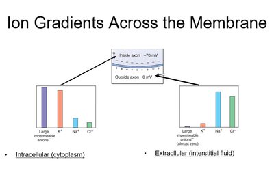
All neurons are electrically excitable, with a resting membrane potential resulting from the distribution of ions across the plasma membrane.
Resting Potential: The voltage difference across the membrane of a neuron not transmitting signals, typically between -40 and -85 mV.
Key Ions: Potassium (K+) and sodium (Na+) are crucial in establishing the resting potential.
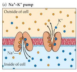
Sodium-Potassium Pump: Uses ATP to move 3 Na+ out and 2 K+ in, maintaining the negative interior.
Leak Channels: Allow K+ to diffuse out, contributing to the negative charge inside.
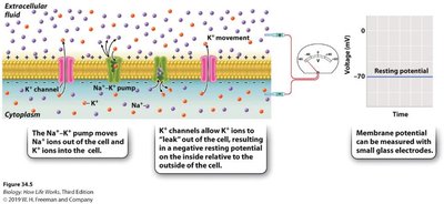
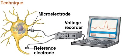
Measuring Membrane Potential
Electrophysiologists use microelectrodes to measure the membrane potential of neurons, providing insights into neuronal excitability and signaling.
Ion Channels and Action Potentials
Types of Ion Channels
Leakage Channels: Always open, contribute to resting potential.
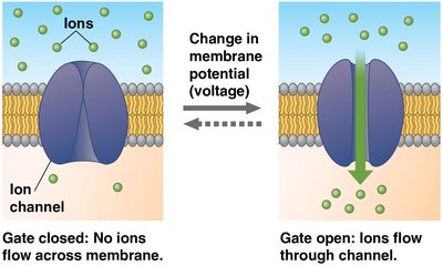
Gated Channels: Open or close in response to stimuli (ligand-gated, voltage-gated, mechanically gated).
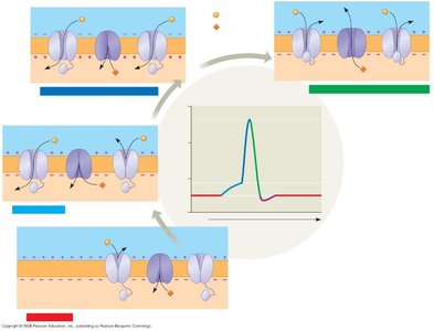
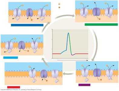
Action Potential Generation
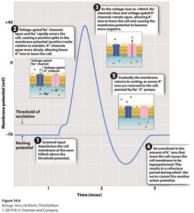
An action potential is a rapid, all-or-none change in membrane potential that propagates along the axon.
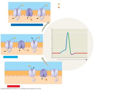
Depolarization: Membrane potential becomes less negative as Na+ enters the cell through voltage-gated channels.
Threshold: If depolarization reaches a critical level (about -50 mV), an action potential is triggered.
Repolarization: Na+ channels close, K+ channels open, and K+ exits, restoring negativity.
Refractory Period: Time during which a neuron cannot fire another action potential, ensuring one-way propagation.

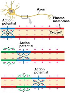
Propagation of Action Potentials
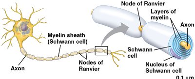
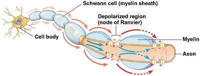
Saltatory Conduction and Myelination
Myelin sheaths, produced by glial cells, insulate axons and increase the speed of action potential propagation. Action potentials jump between nodes of Ranvier in a process called saltatory conduction.
Nodes of Ranvier: Gaps in the myelin sheath where voltage-gated channels are concentrated.
Saltatory Conduction: Rapid transmission of nerve impulses by jumping from node to node.
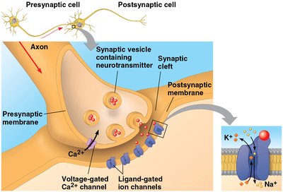
Synaptic Transmission
Chemical and Electrical Synapses
Neurons communicate at synapses, which can be electrical (direct ion flow via gap junctions) or chemical (neurotransmitter-mediated).
Chemical Synapse: Action potential triggers Ca2+ influx, vesicle fusion, and neurotransmitter release into the synaptic cleft.
Neurotransmitter Binding: Opens ligand-gated ion channels on the postsynaptic cell, altering its membrane potential.
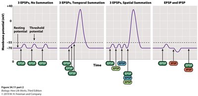
Postsynaptic Potentials and Summation
Excitatory Postsynaptic Potential (EPSP): Depolarizes the postsynaptic membrane, increasing the likelihood of an action potential.
Inhibitory Postsynaptic Potential (IPSP): Hyperpolarizes the membrane, decreasing the likelihood of firing.
Summation: Multiple EPSPs and IPSPs can combine temporally or spatially to determine if threshold is reached.
Neurotransmitters and Receptors
Major Neurotransmitters
Acetylcholine: Involved in muscle stimulation, memory, and learning; acts on both ligand-gated and metabotropic receptors.
Amino Acids: Glutamate (excitatory), glycine, and GABA (inhibitory).
Biogenic Amines: Norepinephrine, dopamine, serotonin—regulate mood, attention, and autonomic functions.
Neuropeptides: Substance P, endorphins—modulate pain perception.
Gases: Nitric oxide (NO), carbon monoxide (CO)—act as local regulators.
Organization of the Nervous System
Central and Peripheral Nervous Systems
Central Nervous System (CNS): Brain and spinal cord; main site of information processing and integration.
Peripheral Nervous System (PNS): Sensory and motor neurons connecting the CNS to the rest of the body.
Somatic Nervous System: Controls voluntary movements.
Autonomic Nervous System: Regulates involuntary functions; divided into sympathetic (fight-or-flight) and parasympathetic (rest-and-digest) divisions.
Sensory Receptors and Signal Transduction
Types of Sensory Receptors
Chemoreceptors: Detect chemical stimuli (e.g., taste, smell).
Mechanoreceptors: Respond to physical deformation (e.g., touch, pressure, sound).
Photoreceptors: Detect light (e.g., rods and cones in the eye).
Sensory Transduction
Sensory receptors convert stimulus energy into changes in membrane potential, initiating action potentials that are interpreted by the CNS as sensations.
Summary Table: Key Ion Channels and Their Functions
Channel Type | Stimulus | Function |
|---|---|---|
Leakage Channel | None (always open) | Maintains resting potential |
Ligand-Gated Channel | Neurotransmitter binding | Postsynaptic potentials (EPSP/IPSP) |
Voltage-Gated Channel | Change in membrane potential | Action potential generation and propagation |
Mechanically Gated Channel | Physical deformation | Touch, pressure, hearing |
Key Equations
Nernst Equation (for equilibrium potential of an ion):
Goldman-Hodgkin-Katz Equation (for membrane potential):
Additional info: This guide covers the foundational concepts of neuron structure, membrane potential, action potential generation, synaptic transmission, and the organization of the nervous system, as well as the roles of glial cells and sensory receptors. These topics are essential for understanding how nervous systems function in animals, as outlined in Campbell Biology in Focus, Chapter 37.
