 Back
BackStep-by-Step Guidance for College Biology Lab Skills and Concepts
Study Guide - Smart Notes
 Tailored notes based on your materials, expanded with key definitions, examples, and context.
Q1. How do you accurately read the volume of a liquid in a graduated cylinder?
Background
Topic: Scientific Measurement – Reading Liquid Volume
This question tests your understanding of how to properly measure the volume of a liquid using a graduated cylinder, a common laboratory tool. Accurate measurement is essential for reliable experimental results in biology and other sciences.
Key Terms and Concepts:
Meniscus: The curved surface of a liquid in a container, caused by surface tension. For most liquids, the correct reading is taken at the bottom of the meniscus.
Graduated Cylinder: A laboratory instrument used to measure the volume of liquids precisely.
Step-by-Step Guidance
Place the graduated cylinder on a flat, level surface to ensure accuracy.
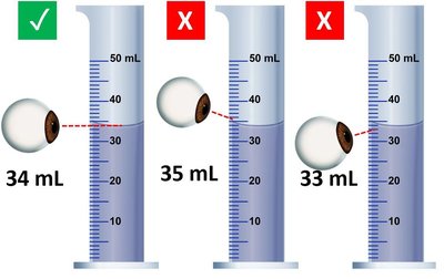
Bring your eyes to the level of the liquid in the cylinder. Do not look from above or below, as this can cause parallax error.
Observe the meniscus. For water and most liquids, read the measurement at the lowest point of the meniscus.
Record the volume at the bottom of the meniscus, aligning your line of sight horizontally with the measurement marking.
Try solving on your own before revealing the answer!
Q2. What are the two major subdivisions of the human skeleton?
Background
Topic: Human Skeletal System
This question assesses your knowledge of the organization of the human skeleton, which is fundamental for understanding anatomy and physiology in biology.
Key Terms:
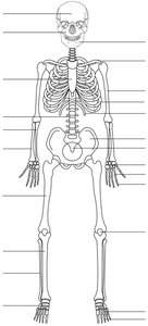
Axial Skeleton: The part of the skeleton that includes the skull, vertebral column, and rib cage.
Appendicular Skeleton: The part of the skeleton that includes the limbs and girdles (shoulder and pelvic girdles).
Step-by-Step Guidance
Recall that the skeleton is divided based on location and function.
Think about which bones protect vital organs and form the central axis of the body.
Consider which bones are involved in movement and are attached to the central skeleton.
Try solving on your own before revealing the answer!
Q3. How do you distinguish between male and female pelvic bones?
Background
Topic: Human Skeletal System – Pelvic Anatomy
This question tests your ability to identify anatomical differences between male and female pelvises, which is important for understanding human biology, reproduction, and forensic science.
Key Terms:
Pelvic Inlet: The upper opening of the pelvis, which is wider in females.
Subpubic Angle: The angle below the pubic symphysis, which is broader in females.
Sacrum: The triangular bone at the base of the spine, which differs in curvature between sexes.
Step-by-Step Guidance
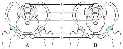
Examine the overall width and shape of the pelvic inlet in both diagrams.
Compare the subpubic angle in each pelvis; a wider angle typically indicates a female pelvis.
Observe the curvature and length of the sacrum and the shape of the pelvic cavity.
Try solving on your own before revealing the answer!
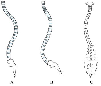
Q4. How do you identify abnormal curvatures of the human spine?
Background
Topic: Human Skeletal System – Vertebral Column
This question focuses on recognizing and naming common vertebral deformities, which is important for understanding musculoskeletal health and clinical conditions.
Key Terms:
Lordosis: Excessive inward curvature of the lumbar spine.
Kyphosis: Excessive outward curvature of the thoracic spine.
Scoliosis: Lateral (sideways) curvature of the spine.
Step-by-Step Guidance
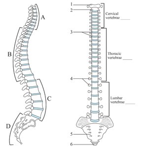
Review the normal curvatures of the spine: cervical and lumbar (lordotic), thoracic and sacral (kyphotic).
Compare the diagrams to identify which one shows an exaggerated lumbar curve (lordosis), an exaggerated thoracic curve (kyphosis), or a lateral deviation (scoliosis).
Match each abnormality to the correct illustration based on the direction and location of the curvature.
Try solving on your own before revealing the answer!
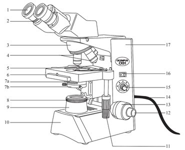
Q5. How do you identify the main parts of a compound microscope?
Background
Topic: Laboratory Equipment – Compound Microscope
This question tests your ability to recognize and label the main components of a compound microscope, which is essential for proper use and maintenance in biological investigations.
Key Terms:
Ocular Lens (Eyepiece): The lens you look through, usually 10x magnification.
Objective Lenses: Lenses of varying magnification (e.g., 4x, 10x, 40x, 100x).
Stage: The platform where the slide is placed.
Coarse and Fine Adjustment Knobs: Used to focus the image.
Light Source: Illuminates the specimen.
Step-by-Step Guidance
Locate the eyepiece at the top of the microscope and note its magnification.
Identify the revolving nosepiece and the attached objective lenses.
Find the stage and the clips or mechanical stage for holding slides.
Locate the coarse and fine adjustment knobs, usually on the side of the microscope.
Identify the light source and diaphragm for adjusting illumination.
Try solving on your own before revealing the answer!
