 Back
BackThe Cell Cycle: Mechanisms and Regulation of Eukaryotic Cell Division
Study Guide - Smart Notes
 Tailored notes based on your materials, expanded with key definitions, examples, and context.
The Cell Cycle
Key Roles of Cell Division
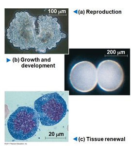
Cell division is a fundamental process in all living organisms, essential for reproduction, growth, tissue renewal, and repair. The continuity of life is based on the ability of cells to divide and produce new cells, ensuring genetic information is faithfully transmitted from one generation to the next.
Reproduction: In unicellular organisms, cell division results in the reproduction of the entire organism.
Growth and Development: In multicellular organisms, cell division enables development from a fertilized cell, growth, and repair of tissues.
Tissue Renewal: Cell division replaces cells that are lost due to normal wear and tear or injury.
Genetic Material Organization
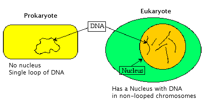
Genetic information in cells is organized into structures called chromosomes, which are composed of DNA and associated proteins. The complete set of genetic material in a cell is known as the genome.
Prokaryotic Cells: Typically have a single, circular DNA molecule and lack a nucleus.
Eukaryotic Cells: Contain multiple, linear DNA molecules housed within a nucleus.
Chromatin, Chromosomes, and Genes
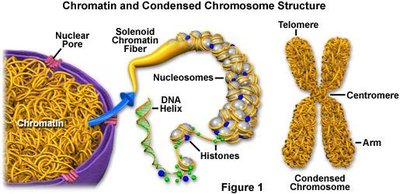
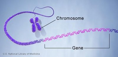
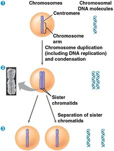
DNA in eukaryotic cells is packaged with proteins to form chromatin. During cell division, chromatin condenses to form visible chromosomes. Each chromosome contains many genes, which are specific sequences of DNA that code for proteins or functional RNA.
Chromatin: The complex of DNA and proteins that is loosely packed in the nucleus during interphase.
Chromosome: A highly condensed structure of chromatin visible during cell division.
Gene: A segment of DNA that encodes a functional product.
Somatic Cells vs. Gametes
Cells in multicellular organisms are classified as either somatic cells or gametes:
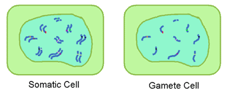
Somatic Cells: Non-reproductive cells with two sets of chromosomes (diploid, 2n).
Gametes: Reproductive cells (sperm and eggs) with one set of chromosomes (haploid, n).
The Mitotic Cell Cycle
Binary Fission in Bacteria
Prokaryotes reproduce by binary fission, a simple form of cell division:
The single, circular chromosome replicates, beginning at the origin of replication.
The two chromosomes move to opposite ends of the cell.
The plasma membrane pinches inward, dividing the cell into two genetically identical daughter cells.
Phases of the Eukaryotic Cell Cycle
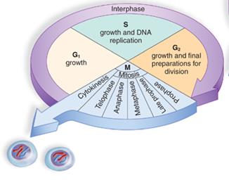
The eukaryotic cell cycle consists of a series of highly regulated phases:
Interphase: Period of cell growth and DNA replication, subdivided into:
G1 phase: Cell growth and normal metabolic activities.
S phase: DNA synthesis and chromosome duplication.
G2 phase: Further growth and preparation for division.
Mitotic (M) Phase: Includes mitosis (nuclear division) and cytokinesis (cytoplasmic division).
Phases of Mitosis
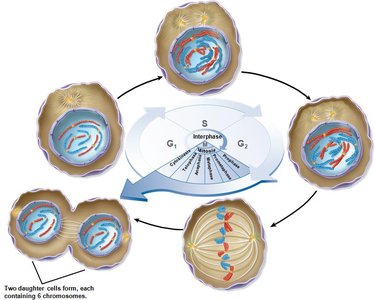
Mitosis is conventionally divided into five phases, ensuring equal distribution of chromosomes to daughter cells:
Prophase: Chromatin condenses into chromosomes; mitotic spindle begins to form; nucleolus disappears.
Prometaphase: Nuclear envelope fragments; spindle microtubules attach to kinetochores on chromosomes.
Metaphase: Chromosomes align at the metaphase plate (center of the cell).
Anaphase: Sister chromatids separate and move toward opposite poles.
Telophase: Chromosomes decondense; nuclear envelopes reform around daughter nuclei.
The Mitotic Spindle
The mitotic spindle is a structure composed of microtubules that orchestrates the movement of chromosomes during mitosis. It consists of:
Centrosomes: Microtubule organizing centers that define the spindle poles.
Microtubules: Three types—astral (position spindle), polar (separate poles), and kinetochore (attach to chromosomes).
Kinetochores: Protein complexes on centromeres where spindle fibers attach.
Cytokinesis
Cytokinesis is the division of the cytoplasm, which usually follows mitosis:
Animal Cells: Occurs by cleavage, forming a cleavage furrow that pinches the cell in two.
Plant Cells: Involves the formation of a cell plate that develops into a new cell wall.
Regulation of the Cell Cycle
Cell Cycle Control System and Checkpoints
The cell cycle is regulated by a molecular control system with checkpoints that ensure each phase is completed accurately before the next begins. The main checkpoints are:
G1 Checkpoint (Restriction Point): Determines if the cell will proceed with division.
G2 Checkpoint: Ensures DNA replication is complete and undamaged.
Metaphase Checkpoint: Verifies that all chromosomes are properly attached to the spindle apparatus.
Checkpoint proteins act as sensors, and loss of checkpoint function can lead to uncontrolled cell division and cancer.
Cyclins, Cdks, and MPF
Progression through the cell cycle is driven by regulatory proteins:
Cyclins: Proteins whose concentrations fluctuate cyclically during the cell cycle.
Cyclin-dependent kinases (Cdks): Enzymes that are active only when bound to cyclins.
MPF (Maturation-Promoting Factor): A cyclin-Cdk complex that triggers passage from G2 to M phase.
Internal and External Signals
Cell cycle progression is influenced by both internal and external signals:
Internal Signals: Unattached kinetochores send signals to delay anaphase until all chromosomes are properly attached.
External Signals: Growth factors (e.g., PDGF) stimulate cell division; density-dependent inhibition and anchorage dependence regulate cell proliferation in tissues.
Loss of Cell Cycle Control and Cancer
Cancer cells evade normal cell cycle controls, leading to uncontrolled proliferation:
May produce their own growth factors or signal without external cues.
Exhibit neither density-dependent inhibition nor anchorage dependence.
Transformation converts a normal cell to a cancerous one; malignant tumors can invade tissues and metastasize.
Summary Table: Comparison of Mitosis and Meiosis
Feature | Mitosis | Meiosis |
|---|---|---|
Number of Divisions | 1 | 2 |
Number of Daughter Cells | 2 | 4 |
Genetic Composition | Identical to parent | Genetically unique |
Chromosome Number | Diploid (2n) | Haploid (n) |
Role | Growth, repair, asexual reproduction | Sexual reproduction, genetic diversity |
Additional info: This table summarizes the key differences between mitosis and meiosis, which are both essential for life but serve distinct biological functions.
