 Back
BackThe Muscular System: Structure, Function, and Contraction Mechanisms
Study Guide - Smart Notes
 Tailored notes based on your materials, expanded with key definitions, examples, and context.
The Muscular System
Introduction to Muscle Tissue
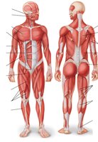
The muscular system is essential for movement, posture, and heat production in the human body. Muscle tissue is specialized for contraction, enabling voluntary and involuntary movements. Muscle cells, also known as muscle fibers, make up approximately 32-40% of body mass and are organized in parallel bundles.
Muscle tissue: Contracts to produce movement.
Voluntary movement: Under conscious control (e.g., skeletal muscle).
Involuntary movement: Not under conscious control (e.g., cardiac and smooth muscle).
Muscles also resist movement and generate heat (about 75% of body heat).
Muscle tissue is excitable, responding to electrical or chemical stimuli.
Types of Muscle Tissue
There are three main types of muscle tissue, each with distinct structure and function:
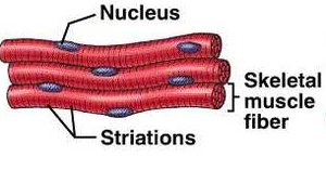
Skeletal muscle: Voluntary, striated, multinucleate, attached to bones for movement.
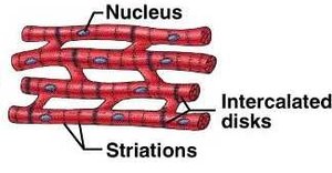
Cardiac muscle: Involuntary, striated, found only in the heart, connected by intercalated disks.
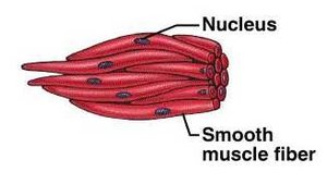
Smooth muscle: Involuntary, non-striated, found in walls of hollow organs (e.g., blood vessels, digestive tract).
Feature | Skeletal Muscle | Cardiac Muscle | Smooth Muscle |
|---|---|---|---|
Striations | Yes | Yes | No |
Control | Voluntary | Involuntary | Involuntary |
Location | Attached to bones | Heart | Walls of organs |
Special Features | Multinucleate | Intercalated disks | Spindle-shaped cells |
Anatomy of Skeletal Muscle
Muscle Structure and Attachments
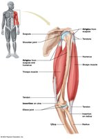
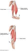
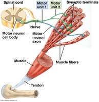
Skeletal muscles are responsible for moving bones and are attached to the skeleton via tendons. Each muscle has an origin (stationary attachment) and an insertion (movable attachment across a joint). Muscles work in groups to coordinate movement:
Synergistic muscles: Work together to produce the same movement.
Antagonistic muscles: Oppose each other's actions.
Gross and Microscopic Structure
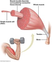
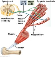
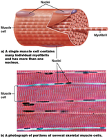
A whole muscle is composed of bundles called fascicles, which are groups of muscle fibers wrapped in connective tissue. Each muscle fiber is a long, multinucleate cell packed with myofibrils, the contractile elements of the cell.
Fascicle: Bundle of muscle fibers.
Myofibril: Rod-like unit within muscle fiber, responsible for contraction.
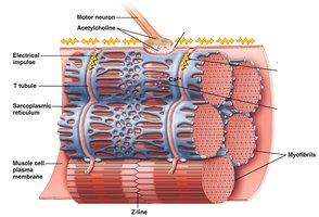
Muscle fibers contain multiple nuclei, mitochondria, sarcoplasmic reticulum, and T-tubules.
Sarcomere Structure
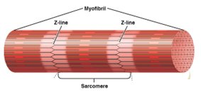
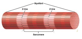
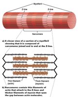
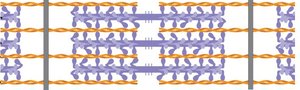
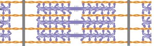
The sarcomere is the basic contractile unit of a myofibril, defined by Z lines at each end. Sarcomeres contain thick (myosin) and thin (actin) filaments, which interact during muscle contraction.
Z lines: Boundaries of a sarcomere.
M line: Center of the sarcomere.
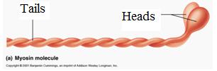
Thick filaments: Composed of myosin.
Thin filaments: Composed of actin, tropomyosin, and troponin complex.
Muscle Contraction Mechanisms
Neuromuscular Junction and Action Potential
Muscle contraction is initiated by signals from motor neurons. The neuromuscular junction is the synapse where a motor neuron communicates with a muscle fiber. The neurotransmitter acetylcholine (Ach) is released, triggering an action potential in the muscle cell membrane.
Acetylcholine (Ach): Neurotransmitter that initiates muscle contraction.
Action potential (AP): Electrical impulse that travels along the muscle fiber membrane.
AP triggers the release of calcium ions (Ca2+) from the sarcoplasmic reticulum.
The Sliding Filament Theory
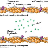
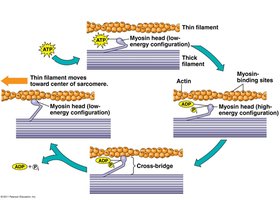
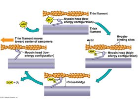
The sliding filament theory explains how muscles contract at the molecular level. When Ca2+ binds to the troponin complex, it causes a conformational change that moves tropomyosin, exposing the active sites on actin. Myosin heads bind to these sites, forming cross-bridges, and use ATP to pull the thin filaments toward the center of the sarcomere, shortening the muscle.
At rest, myosin heads are not attached to actin; active sites are blocked by tropomyosin.
Ca2+ binding to troponin exposes the active sites.
Myosin heads attach, perform a powerstroke, and detach when new ATP binds.
This cycle repeats as long as Ca2+ and ATP are present.
Energy for Muscle Contraction
Muscle contraction requires large amounts of ATP, which is supplied by several sources:
Creatine phosphate: Rapidly donates phosphate to ADP to regenerate ATP.
Stored glycogen: Broken down to glucose for ATP production.
Other molecules: Fats and proteins can also be used for ATP synthesis if necessary.
Muscle fibers are either fully contracted or relaxed (all-or-none principle). A muscle twitch is a single contraction-relaxation cycle. Summation occurs when stimuli are frequent enough to prevent relaxation, increasing force. Tetanus is sustained maximal contraction.
Muscle Fiber Types and Exercise
Muscle fibers can be classified based on contraction speed and endurance:
Slow-twitch fibers: Contract slowly, resist fatigue, suited for endurance activities.
Fast-twitch fibers: Contract quickly, fatigue rapidly, suited for short bursts of power.
Most muscles contain a mix of both types.
Exercise improves muscle tone, strength, and endurance. Strength training increases muscle mass (not cell number), while aerobic training enhances endurance and mitochondrial content.
Other Types of Muscle
Cardiac Muscle
Cardiac muscle is found only in the heart. It is striated, involuntary, and contracts at a moderate speed without fatigue. Cells are connected by intercalated disks and gap junctions, allowing action potentials to spread rapidly.
Smooth Muscle
Smooth muscle is found in the walls of blood vessels and hollow organs. It contracts slowly, is not striated, and does not fatigue easily. It is regulated by the autonomic nervous system.
Diseases of the Muscular System
Muscular Dystrophy
Muscular dystrophy refers to a group of genetic diseases causing progressive weakness and degeneration of skeletal muscles. Duchenne muscular dystrophy is the most common form. Muscle fibers are damaged, leading to Ca2+ influx and protein destruction.
Tetanus
Tetanus is caused by infection with Clostridium tetani bacteria, which produce a toxin that causes sustained muscle contraction. It is preventable by vaccination.
Summary Table: Muscle Types
Muscle Type | Striations | Control | Location | Special Features |
|---|---|---|---|---|
Skeletal | Yes | Voluntary | Attached to bones | Multinucleate, rapid contraction |
Cardiac | Yes | Involuntary | Heart | Intercalated disks, rhythmic contraction |
Smooth | No | Involuntary | Walls of organs | Spindle-shaped, slow contraction |
Additional info: The sliding filament theory and the role of ATP in muscle contraction are foundational concepts for understanding muscle physiology. Disorders such as muscular dystrophy and tetanus highlight the importance of proper muscle function and the consequences of its disruption.
