 Back
BackBio 100 LEC Chapter 5 Part 2 UPDATED
Study Guide - Smart Notes
 Tailored notes based on your materials, expanded with key definitions, examples, and context.
Bio 100 LEC Chapter 5 Part 2
Chapter 5: The Structure and Function of Large Biological Molecules
Module 4: Proteins
This module explores the structure, diversity, and function of proteins, one of the four major classes of large biological molecules. Proteins are essential to nearly every process within living cells, and their structure is intricately linked to their function.
Concept 5.4: Proteins—Diversity of Structure and Function
Categories and Functions of Proteins
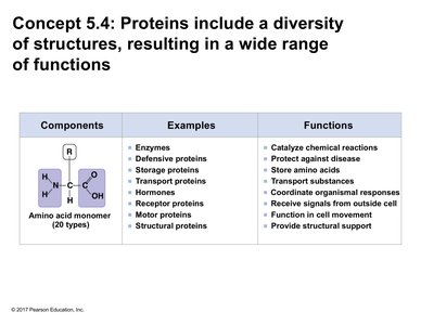
Proteins account for more than 50% of the dry mass of most cells and are involved in nearly every cellular function. Their diverse structures allow for a wide range of functions, which can be grouped into several categories:
Component | Examples | Functions |
|---|---|---|
Amino acid monomer (20 types) |
|
|
Amino Acids: The Building Blocks of Proteins
Structure of Amino Acids
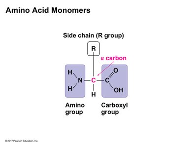
An amino acid is an organic molecule with a central (alpha) carbon atom bonded to four different groups: an amino group, a carboxyl group, a hydrogen atom, and a variable side chain (R group). The R group determines the unique characteristics of each amino acid.
At physiological pH, the amino group is typically protonated (NH3+) and the carboxyl group is deprotonated (COO-).
The properties of the R group confer the chemical behavior of the amino acid.
Classification of Amino Acids by Side Chain Properties
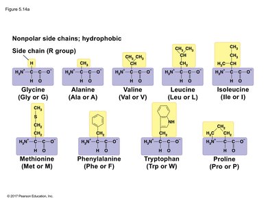
Nonpolar (Hydrophobic) Side Chains
Nonpolar amino acids have side chains that are primarily hydrocarbons. These amino acids tend to cluster in the interior of proteins, away from water, and are abundant in membrane-spanning regions.
Polar (Hydrophilic) Side Chains
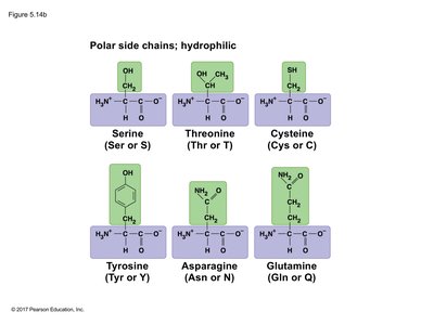
Polar amino acids have side chains that can form hydrogen bonds with water, making them hydrophilic. These residues are often found on the exterior of proteins, interacting with the aqueous environment.
Electrically Charged (Hydrophilic) Side Chains
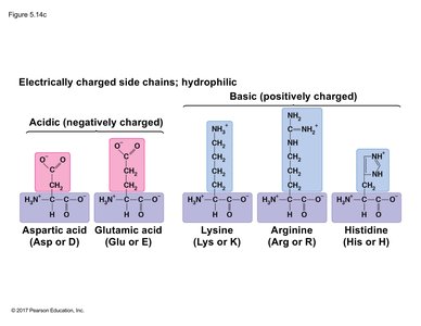
Some amino acids have side chains that are charged at physiological pH. Acidic side chains are negatively charged, while basic side chains are positively charged. These residues can form ionic bonds and participate in interactions critical for protein structure and function.
Polypeptides: Formation and Structure
Peptide Bond Formation
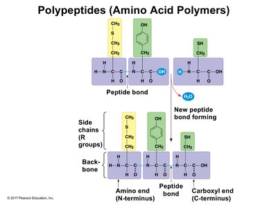
Proteins are polymers of amino acids, linked by peptide bonds formed through condensation (dehydration synthesis) reactions. Each polypeptide has directionality, with an N-terminus (amino end) and a C-terminus (carboxyl end).
Peptide bonds form between the carboxyl group of one amino acid and the amino group of the next.
The sequence of amino acids (primary structure) determines the protein's final shape and function.
Protein Structure and Function
Relationship Between Structure and Function
The specific activities of proteins result from their intricate three-dimensional architecture. The folding and shape of a protein are essential for its biological activity.
Visualizing Protein Structure
Models of Protein Structure
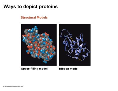
Proteins can be depicted using various models, each highlighting different aspects:
Space-filling model: Shows the spatial arrangement of atoms.
Ribbon model: Emphasizes the backbone and folding patterns (e.g., alpha helices, beta sheets).
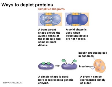
Simplified Diagrams
Simplified diagrams may show only the overall shape or represent proteins as simple shapes or dots, depending on the context (e.g., enzyme-substrate interactions, hormone secretion).
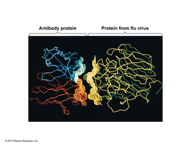
Protein-Protein Interactions
Proteins often interact with other proteins, such as antibodies binding to viral proteins. The complementarity of their shapes is crucial for these interactions.
Levels of Protein Structure
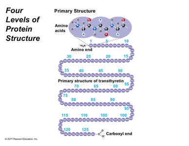
Primary Structure
The primary structure is the unique sequence of amino acids in a polypeptide, held together by peptide bonds. This sequence dictates all higher levels of structure.
Secondary Structure
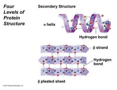
The secondary structure consists of regular coils and folds stabilized by hydrogen bonds between backbone atoms. The two main types are:
Alpha helix (α-helix): A spiral structure stabilized by hydrogen bonds within the same polypeptide chain.
Beta pleated sheet (β-sheet): Sheet-like structures formed by hydrogen bonds between segments of the polypeptide chain, which may be parallel or antiparallel.
Tertiary Structure
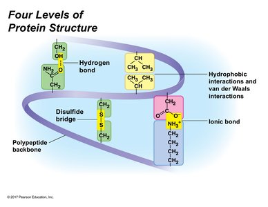
The tertiary structure is the overall three-dimensional shape of a polypeptide, resulting from interactions between R groups (side chains). These include:
Hydrogen bonds
Ionic bonds
Hydrophobic interactions
Van der Waals interactions
Disulfide bridges (covalent bonds between cysteine residues)
Quaternary Structure
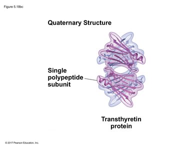
The quaternary structure arises when two or more polypeptide chains (subunits) assemble into a functional protein. The same types of interactions that stabilize tertiary structure also stabilize quaternary structure.
Protein Denaturation and Renaturation
Effects of Environmental Conditions
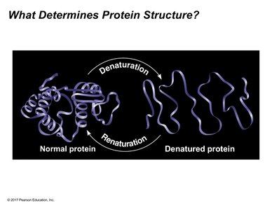
Protein structure can be disrupted by changes in pH, temperature, or salinity, leading to denaturation—the loss of native structure and function. Under certain conditions, some proteins can refold (renature) and regain function.
Case Study: Hemoglobin and Sickle-Cell Disease
Hemoglobin Structure
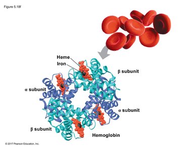
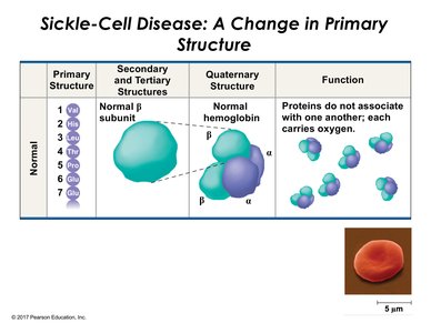
Hemoglobin is a tetrameric protein (four subunits: two alpha and two beta) found in red blood cells, responsible for oxygen transport.
Sickle-Cell Disease: A Change in Primary Structure
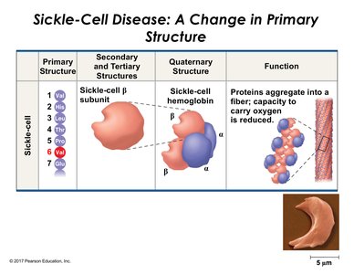
Sickle-cell disease is caused by a single amino acid substitution (glutamic acid to valine) in the beta subunit of hemoglobin. This change alters the protein's properties, causing hemoglobin molecules to aggregate and distort red blood cell shape, impairing oxygen transport.
Primary Structure | Secondary and Tertiary Structures | Quaternary Structure | Function |
|---|---|---|---|
Normal: Glu at position 6 | Normal β subunit | Normal hemoglobin | Efficient oxygen transport |
Sickle-cell: Val at position 6 | Sickle β subunit | Sickle hemoglobin (aggregates) | Impaired oxygen transport |
Determining Protein Structure
X-ray Crystallography
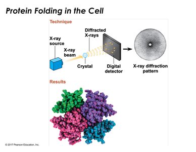
X-ray crystallography is a key technique for determining the three-dimensional structure of proteins. It involves crystallizing the protein, exposing it to X-rays, and analyzing the diffraction pattern to deduce atomic positions.
Additional info: Understanding protein structure is fundamental to biochemistry, molecular biology, and medicine, as it underpins enzyme function, cellular signaling, and the molecular basis of many diseases.
