Pearson+ LogoPearson+ Logo
Ch. 12 - The Cell Cycle
Freeman - Biological Science 7th Edition
Freeman7th EditionBiological ScienceISBN: 9783584863285Not the one you use?Change textbook
Chapter 12, Problem 6

What evidence suggests that during anaphase, kinetochore microtubules shorten at the kinetochore?

Verified step by step guidance
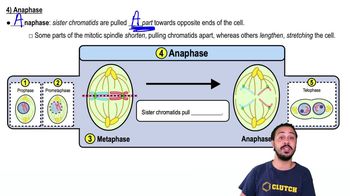
1
Understand the structure and function of kinetochore microtubules: Kinetochore microtubules are specialized structures that attach chromosomes to the spindle apparatus during cell division. They play a crucial role in ensuring accurate chromosome segregation.
Review the process of anaphase: During anaphase, sister chromatids separate and are pulled towards opposite poles of the cell. This movement is facilitated by the shortening of kinetochore microtubules.
Examine fluorescence microscopy evidence: Fluorescence microscopy has been used to tag kinetochore microtubules with fluorescent markers. Observations show that the fluorescent markers at the kinetochores move toward the spindle poles, indicating that the microtubules are shortening at the kinetochore end.
Consider the role of motor proteins: Motor proteins such as dynein and kinesin are known to be involved in the movement of chromosomes. These proteins can walk along microtubules, and their activity at the kinetochore can contribute to the shortening process.
Evaluate biochemical experiments: Biochemical experiments that manipulate the activity of motor proteins or the stability of microtubules provide further evidence. For example, drugs that stabilize microtubules can inhibit chromosome movement, supporting the idea that microtubule disassembly at the kinetochore is necessary for chromosome segregation.

Verified video answer for a similar problem:

This video solution was recommended by our tutors as helpful for the problem above.
Video duration:
2m
Was this helpful?

Key Concepts

Here are the essential concepts you must grasp in order to answer the question correctly.

Anaphase

Anaphase is a stage in mitosis where sister chromatids are pulled apart towards opposite poles of the cell. This process is crucial for ensuring that each daughter cell receives an identical set of chromosomes. During anaphase, the cohesin proteins that hold the sister chromatids together are cleaved, allowing them to separate and move due to the action of the spindle apparatus.
Recommended video:

Kinetochore Microtubules

Kinetochore microtubules are specialized structures that connect the spindle apparatus to the kinetochore, a protein complex assembled on the centromere of each chromosome. These microtubules play a vital role in chromosome movement during cell division by exerting forces that pull the chromatids apart. The dynamic nature of these microtubules allows them to grow and shrink, facilitating the movement of chromosomes.
Recommended video:

Microtubule Shortening

Microtubule shortening refers to the process where the length of microtubules decreases, which is essential for the movement of chromosomes during anaphase. This shortening occurs primarily at the kinetochore, where depolymerization of tubulin subunits takes place. Evidence for this process includes live-cell imaging studies that show the rapid reduction in microtubule length as chromatids are pulled towards the poles, indicating that the force required for movement is generated by this shortening.
Recommended video:
Guided course
12:51
Spermatogenesis