 Back
BackCell Biology: Membranes, Cytoskeleton, and Protein Trafficking Study Guide
Study Guide - Smart Notes
 Tailored notes based on your materials, expanded with key definitions, examples, and context.
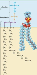
Q1. Which of the structures represented above is/are components(s) of a biological membrane?
Background
Topic: Biological Membranes
This question tests your understanding of the molecular components that make up biological membranes, such as phospholipids, cholesterol, and proteins.
Key Terms:
Phospholipids: Major structural component of membranes, forming the lipid bilayer.
Cholesterol: Modulates membrane fluidity.
Proteins: Embedded or associated with the membrane, serving various functions.
Step-by-Step Guidance
Examine the molecular structures labeled A, B, C, and D in the provided image. Identify which ones are phospholipids, cholesterol, or other membrane-associated molecules.
Recall that biological membranes are primarily composed of a phospholipid bilayer, with cholesterol and proteins interspersed.
Determine which structures are found in the membrane based on their chemical features (e.g., hydrophobic tails, hydrophilic heads for phospholipids).
Try solving on your own before revealing the answer!
Final Answer: A, B, and D
Structures A and B are phospholipids and cholesterol, both of which are membrane components. D is likely a glycolipid or another membrane-associated molecule. C is not a typical membrane component.
Q2. Which component of a phospholipid is found in the interior of a lipid bilayer?
Background
Topic: Membrane Structure
This question tests your knowledge of the amphipathic nature of phospholipids and how they arrange themselves in a bilayer.
Key Terms:
Hydrophobic: Water-fearing, nonpolar (fatty acid tails).
Hydrophilic: Water-loving, polar (glycerol and phosphate group).
Step-by-Step Guidance
Recall the structure of a phospholipid: a glycerol backbone, two fatty acid tails, and a phosphate group.
Understand that in a bilayer, hydrophobic tails face inward, away from water, while hydrophilic heads face outward.
Identify which component (glycerol, fatty acids, or phosphate group) is located in the interior of the bilayer.
Try solving on your own before revealing the answer!
Final Answer: Fatty acids
The fatty acid tails are hydrophobic and are found in the interior of the lipid bilayer, away from the aqueous environment.
Q3. Which of the molecules above maintain membrane fluidity at low temperatures and keep membranes from becoming too fluid at higher temperatures?
Background
Topic: Membrane Fluidity
This question tests your understanding of how cholesterol and other molecules regulate the fluidity of cell membranes.
Key Terms:
Cholesterol: A lipid that modulates membrane fluidity by preventing tight packing at low temperatures and restricting movement at high temperatures.
Step-by-Step Guidance
Identify which molecule in the image is cholesterol.
Recall cholesterol's dual role in maintaining membrane fluidity across temperature changes.
Match the correct molecule to the function described in the question.
Try solving on your own before revealing the answer!
Final Answer: B
Cholesterol is responsible for maintaining membrane fluidity at different temperatures.
Q4. Which of the following is not part of the endomembrane system?
Background
Topic: Endomembrane System
This question tests your knowledge of the organelles that make up the endomembrane system in eukaryotic cells.
Key Terms:
Endomembrane system: Includes the nuclear envelope, endoplasmic reticulum, Golgi apparatus, lysosomes, vesicles, and plasma membrane.
Chloroplasts: Organelles involved in photosynthesis, not part of the endomembrane system.
Step-by-Step Guidance
List the components of the endomembrane system.
Identify which option is not included in this system.
Try solving on your own before revealing the answer!
Final Answer: Chloroplasts
Chloroplasts are not part of the endomembrane system; they are involved in photosynthesis.
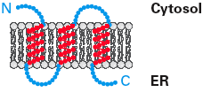
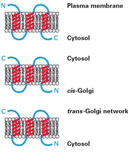
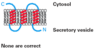
Q5. A transmembrane protein was synthesized in the orientation shown to the left (N = amino- and C = carboxy- terminus). Which of the following accurately depict(s) the orientation of this protein as it travels to the plasma membrane?
Background
Topic: Protein Trafficking and Membrane Topology
This question tests your understanding of how membrane proteins maintain their orientation as they move through the endomembrane system to the plasma membrane.
Key Terms:
Transmembrane protein: Spans the lipid bilayer with specific orientation of N- and C-termini.
Endoplasmic reticulum (ER): Site of protein synthesis and insertion into the membrane.
Vesicular transport: Proteins are trafficked in vesicles, maintaining their orientation.
Step-by-Step Guidance
Examine the initial orientation of the protein in the ER membrane (N-terminus in cytosol or lumen).
Recall that during vesicular transport, the topology of the protein is preserved (cytosolic side remains cytosolic, lumenal side remains lumenal).
Compare the options provided in the images to determine which one maintains the correct orientation as the protein reaches the plasma membrane.
Try solving on your own before revealing the answer!
Final Answer: The orientation where the N-terminus remains on the cytosolic side and the C-terminus on the extracellular side (as shown in the plasma membrane diagram).
This is because the topology established in the ER is maintained throughout the secretory pathway.
Q6. All of the following statements are true for microtubules and microfilaments, EXCEPT:
Background
Topic: Cytoskeleton
This question tests your ability to distinguish between the properties and functions of microtubules and microfilaments (actin filaments).
Key Terms:
Microtubules: Hollow tubes made of tubulin, involved in cell shape, division, and transport.
Microfilaments: Thin filaments made of actin, involved in cell movement and structure.
MTOC (Microtubule Organizing Center): Structure from which microtubules originate.
Step-by-Step Guidance
Review each statement and recall the unique features of microtubules and microfilaments.
Identify which statement does not apply to both filament types (e.g., origin from MTOC is specific to microtubules).
Try solving on your own before revealing the answer!
Final Answer: Both filaments originate from structure known as MTOC (False for microfilaments).
Microfilaments do not originate from the MTOC; only microtubules do.
Q7. A microtubule protofilament is formed by the:
Background
Topic: Microtubule Structure
This question tests your understanding of how microtubules are assembled from tubulin subunits.
Key Terms:
Tubulin dimer: Composed of alpha- and beta-tubulin, building block of microtubules.
Protofilament: Linear chain of tubulin dimers.
Step-by-Step Guidance
Recall that microtubules are made of protofilaments, which are linear chains of tubulin dimers.
Determine whether the association is lateral or head-to-tail, and which tubulin subunit is involved.
Try solving on your own before revealing the answer!
Final Answer: Head-to-tail association of tubulin dimers
Protofilaments are formed by the head-to-tail association of alpha- and beta-tubulin dimers.
Q8. In the process called actin filaments treadmilling:
Background
Topic: Actin Dynamics
This question tests your understanding of the dynamic behavior of actin filaments, specifically treadmilling.
Key Terms:
Treadmilling: Process where actin subunits are added at the + end and removed from the - end, causing the filament to appear to "move" without changing length.
Step-by-Step Guidance
Recall the definition of treadmilling and how it powers cellular movements.
Identify which statement best describes this process (e.g., subunit addition/removal, role in movement).
Try solving on your own before revealing the answer!
Final Answer: This process can power intracellular movements.
Treadmilling allows actin filaments to drive movement within cells.
Q9. Which of the following is not included in coated budding vesicle?
Background
Topic: Vesicular Transport
This question tests your knowledge of the components involved in the formation of coated vesicles during intracellular transport.
Key Terms:
Coat proteins: Help shape the vesicle and select cargo.
GTPase switch proteins: Regulate vesicle formation and uncoating.
t-SNAREs: Involved in vesicle fusion, not budding.
Step-by-Step Guidance
List the components involved in vesicle budding (coat proteins, GTPases, cargo, sorting signals).
Identify which component is involved in vesicle fusion rather than budding.
Try solving on your own before revealing the answer!
Final Answer: t-SNAREs
t-SNAREs are involved in vesicle fusion, not in the budding process.
Q10. Vesicle coat functions in selecting specific proteins for cargo
Background
Topic: Vesicular Transport
This question tests your understanding of the role of vesicle coats in cargo selection during vesicle formation.
Key Terms:
Vesicle coat: Protein layer that helps shape the vesicle and select cargo proteins.
Step-by-Step Guidance
Recall the function of vesicle coats (e.g., clathrin, COPI, COPII) in cargo selection and vesicle formation.
Determine whether the statement is true or false based on your knowledge.
Try solving on your own before revealing the answer!
Final Answer: True
Vesicle coats do select specific proteins for cargo during vesicle formation.
Q11. Centrioles are examples of doublet microtubules
Background
Topic: Microtubule Structure
This question tests your knowledge of the structure of centrioles and the types of microtubules they contain.
Key Terms:
Centrioles: Cylindrical structures composed of microtubule triplets, not doublets.
Doublet microtubules: Found in cilia and flagella, not centrioles.
Step-by-Step Guidance
Recall the arrangement of microtubules in centrioles (triplets) versus cilia/flagella (doublets).
Determine whether the statement is true or false based on this structural difference.
Try solving on your own before revealing the answer!
Final Answer: False
Centrioles are composed of microtubule triplets, not doublets.
