 Back
BackCell Biology Study Guide: Plasma Membrane Structure, Function, and Aerobic Respiration
Study Guide - Smart Notes
 Tailored notes based on your materials, expanded with key definitions, examples, and context.
The Structure and Function of the Plasma Membrane
Overview of Plasma Membrane
The plasma membrane is a thin, fragile structure (5–10 nm thick) that separates the cell from its external environment. It is essential for maintaining cellular integrity and regulating interactions with the surroundings.
Compartmentalization: Membranes enclose the cell and intracellular spaces, allowing specialized activities to occur independently.
Scaffold for Biochemical Activities: Membranes serve as platforms for organizing enzymes and facilitating metabolic pathways.
Selectively Permeable Barrier: The membrane acts as a barrier, controlling the entry and exit of substances.
Transporting Solutes: Specialized transport machinery moves ions, sugars, and amino acids across the membrane.
Response to External Signals: Membranes play a critical role in signal transduction, responding to hormones and growth factors.
Intercellular Interactions: Membranes allow cells to recognize and signal one another.
Energy Transduction: Membranes are involved in converting energy, as seen in mitochondria and chloroplasts.
Membrane Structure: Lipid Bilayer
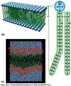
The plasma membrane is primarily composed of a lipid bilayer, with hydrophilic heads facing outward and hydrophobic tails inward.
Lipid Bilayer: Proposed by Gorter & Grendel (1925), the bilayer consists of two layers of lipids with hydrophilic heads exposed to aqueous environments.
Chemical Composition of Membranes
Membranes are lipid-protein assemblies held together by noncovalent bonds. The lipid:protein ratio varies by membrane type, organism, and cell type.
Membrane Lipids: Amphipathic molecules with hydrophobic and hydrophilic regions.
Three Main Types: Phosphoglycerides, sphingolipids, and cholesterol.
Phosphoglycerides
Most membrane phospholipids are built on a glycerol backbone and are diglycerides, with two fatty acids esterified to glycerol. 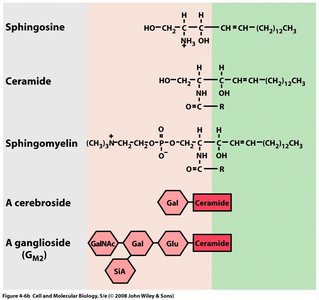
Sphingolipids
Sphingolipids are derivatives of sphingosine, an amino alcohol with a long hydrocarbon chain. Ceramide is formed by linking sphingosine to a fatty acid. Sphingolipids include sphingomyelin, cerebrosides, and gangliosides. 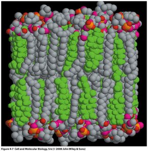
Cholesterol
Cholesterol is a sterol present in animal membranes, absent in most plant and all bacterial membranes. Its rigid ring structure interferes with phospholipid tail movement, affecting membrane fluidity.
Membrane Carbohydrates
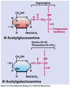
Eukaryotic plasma membranes contain carbohydrates, which are covalently linked to lipids (glycolipids) or proteins (glycoproteins).
Orientation: All membrane carbohydrates face outward, either toward the extracellular space or organelle interior.
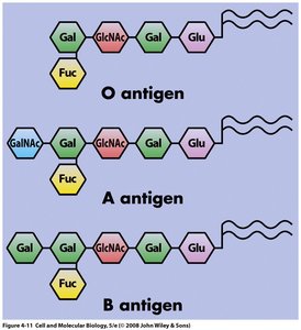
Glycolipids: Short, branched oligosaccharide chains; determine ABO blood type and serve as receptors for certain pathogens.
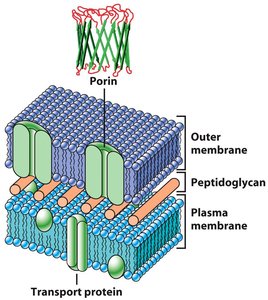
Structure and Function of Membrane Proteins
Classes of Membrane Proteins
Membrane proteins are classified by their relationship to the lipid bilayer:
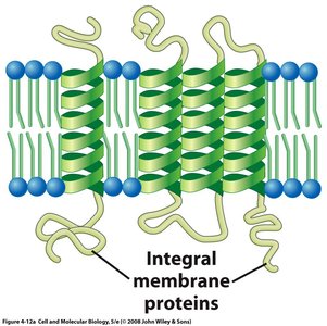
Integral Proteins: Penetrate the bilayer, often spanning it completely (transmembrane proteins).
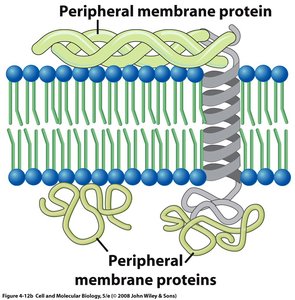
Peripheral Proteins: Located outside the bilayer, attached by weak electrostatic bonds to lipid head groups or integral proteins.
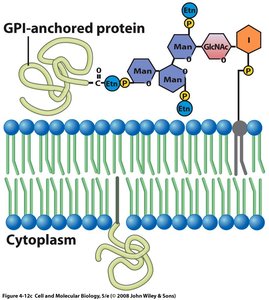
Lipid-Anchored Proteins: Covalently linked to membrane lipids, exposed on either side of the membrane.
Functions of Membrane Proteins
Receptors: Bind specific substances at the membrane surface.
Channels/Transporters: Facilitate movement of ions and solutes.
Electron Transfer: Participate in photosynthesis and respiration.
Identifying Transmembrane Segments
Hydropathy plots are used to identify transmembrane segments by measuring the hydrophobicity of amino acid sequences. Peaks in hydrophobicity indicate likely membrane-spanning regions. 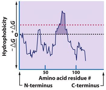
Movement of Substances Across Cell Membranes
Diffusion and Osmosis
The plasma membrane retains dissolved materials and allows exchange of substances.
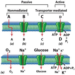
Passive Movement: Includes simple diffusion through the lipid bilayer or protein-lined channels.
Active Movement: Requires energy to move substances against concentration gradients.
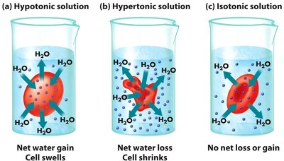
Osmosis
Water moves through semipermeable membranes from regions of lower solute concentration to higher solute concentration.
Hypotonic: Cell swells due to net water gain.
Hypertonic: Cell shrinks due to net water loss.
Isotonic: No net water movement.
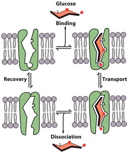
Facilitated Diffusion
Facilitated diffusion involves a membrane-spanning protein (facilitative transporter) that selectively binds a substance and changes shape to allow its movement across the membrane.
Transporter: Binds solute on one side, undergoes conformational change, and releases solute on the other side.
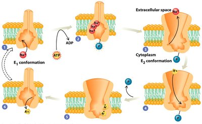
Active Transport
Active transport generates strong ion gradients by expending energy, often coupled to ATP hydrolysis.
Na+/K+ Pump: Pumps 3 Na+ ions out and 2 K+ ions in per cycle, maintaining cell volume and electrochemical gradients.
P-type Ion Pump: Requires phosphorylation during the cycle, altering affinity for ions and exposing binding sites to different membrane sides.
Aerobic Respiration and the Mitochondrion
Introduction to Aerobic Respiration
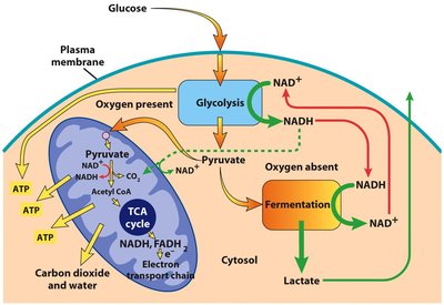
Aerobic respiration is the process by which eukaryotic cells extract energy from organic molecules using oxygen, primarily in the mitochondrion.
Early Earth: Populated by anaerobes; aerobes evolved after oxygen accumulation.
Mitochondria: Site of aerobic respiration in eukaryotes.
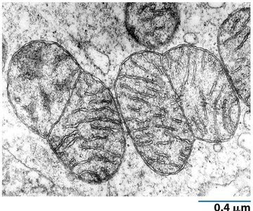
Mitochondrial Structure and Function
Mitochondria are bean-shaped organelles with two membranes: an outer membrane and a highly folded inner membrane (cristae).
Outer Membrane: Contains porin, permeable to small molecules.
Inner Membrane: Impermeable, contains cardiolipin, and is the site of ATP synthesis.
Matrix: Contains DNA, ribosomes, enzymes, and is the site of RNA/protein synthesis and heme group synthesis.
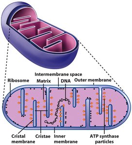
Mitochondrial Membranes
Outer Membrane: Permeable, contains porin.
Inner Membrane: Impermeable, contains cardiolipin and diphosphatidylglycerol.
Mitochondrial Matrix
Contents: Circular DNA, ribosomes, enzymes, high protein concentration.
Functions: RNA/protein synthesis, Ca2+ transport, heme synthesis.
Oxidative Metabolism in the Mitochondrion
Glycolysis: Occurs in the cytosol, produces NADH and pyruvate.
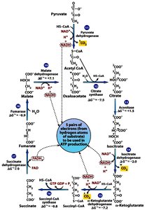
TCA Cycle: Occurs in the mitochondrial matrix, produces NADH, FADH2, and ATP.
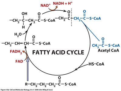
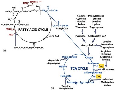
Fatty Acid Cycle and TCA Cycle Connection
Fatty acids are broken down to acetyl CoA, which enters the TCA cycle.
HS-CoA: Removes one carbon at a time from fatty acids.
Interactions Between Cells and Their Environment
Extracellular Space and Matrix
Glycocalyx: Mediates cell-cell interactions, provides mechanical protection.
Extracellular Matrix (ECM): Organized network of extracellular materials, regulates cell shape and activities.
Components of the ECM
Collagens: Fibrous glycoproteins, provide tensile strength, most abundant protein in the human body.
Proteoglycans: Protein-polysaccharide complexes, attract water and form hydrated gels.
Fibronectin: Modular protein, links ECM components.
Laminin: Influences cell migration, growth, and differentiation.
Entactin: Component of basement membranes, aids adhesion and penetration.
Cell-ECM Interactions: Integrins
Integrins are integral membrane proteins that mediate adhesion to the substratum and signal transmission.
Structure: Composed of two subunits (α and β).
Function: Adhesion and signal transduction.
Cell-Cell Adhesion: Junctions
Adherens Junctions: Ca2+-dependent, common in epithelia.
Desmosomes: Disk-shaped, numerous in tissues under mechanical stress.
Tight Junctions: Seal adjacent cells, control paracellular transport.
Gap Junctions: Allow intercellular communication, composed of connexins.
Membrane Component | Structure | Function |
|---|---|---|
Phosphoglycerides | Glycerol backbone, two fatty acids | Structural, amphipathic |
Sphingolipids | Sphingosine backbone, fatty acid | Structural, signaling |
Cholesterol | Sterol ring, hydroxyl group | Fluidity regulation |
Integral Proteins | Transmembrane domains | Transport, signaling |
Peripheral Proteins | Noncovalent attachment | Support, enzymatic |
Lipid-Anchored Proteins | Covalent lipid linkage | Signaling, adhesion |
Transport Type | Energy Requirement | Direction |
|---|---|---|
Simple Diffusion | No | Down gradient |
Facilitated Diffusion | No | Down gradient |
Active Transport | Yes (ATP) | Against gradient |
Additional info: Academic context was added to clarify the structure and function of membrane components, transport mechanisms, and mitochondrial processes. Tables were inferred to summarize key comparisons and classifications.
