 Back
BackCell Types and Membrane Structure: Foundations of Cell Biology
Study Guide - Smart Notes
 Tailored notes based on your materials, expanded with key definitions, examples, and context.
Cell Types: Prokaryotic vs. Eukaryotic
Overview of Basic Cell Types
Cells are the fundamental units of life, and all organisms are composed of cells. There are two primary cell types: prokaryotic and eukaryotic. Both types share certain features, such as a cell membrane, cytoplasm, and genetic material, but they differ significantly in their internal structure and complexity.
Prokaryotic cells: Found in bacteria; lack membrane-bound organelles and a defined nucleus.
Eukaryotic cells: Include plants, animals, fungi, protozoa, and yeast; possess membrane-bound organelles and a nucleus.
Common features: Cell membrane, cytoplasm, chromosomes (DNA), ribosomes, cell wall (in many cases), flagella (structure and movement differ).
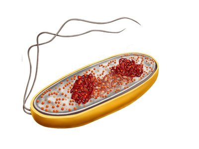
Prokaryotic Cell Structure
Prokaryotic cells are characterized by their simplicity and lack of compartmentalization. Their genetic material is located in a region called the nucleoid, and they often possess a capsule (slime layer) for protection and adhesion.
Capsule functions: Food storage, surface adhesion, increased pathogenicity, protection from desiccation.
Cell wall: Determines cell shape and protects against osmotic changes.
Shapes of Prokaryotic Cells
Bacterial cell walls determine the shape of the cell, which can be classified into three main types:
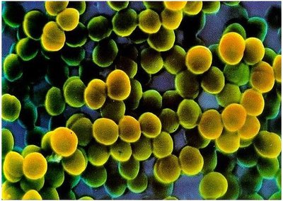
Cocci: Spherical bacteria.
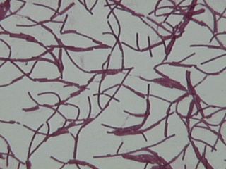
Bacilli: Rod-shaped bacteria.
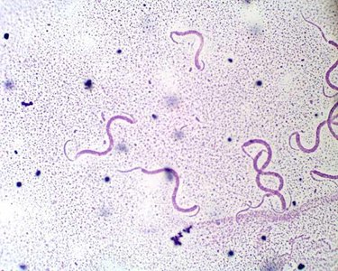
Spirilli: Spiral-shaped bacteria.
Eukaryotic Cell Structure
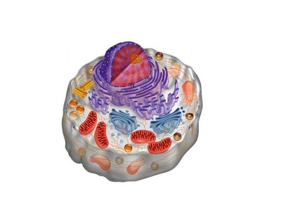
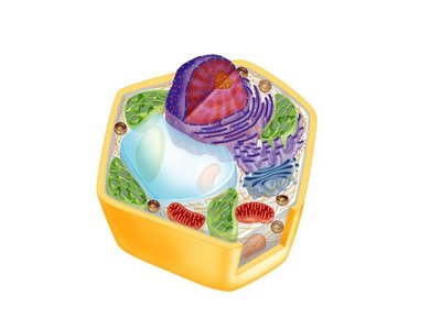
Eukaryotic cells are more complex, containing numerous organelles that perform specialized functions. Both plant and animal cells are eukaryotic, but they have distinct features.
Animal cells: Contain centrioles, lysosomes, mitochondria, endoplasmic reticulum, Golgi apparatus, peroxisomes, and cytoskeletal elements.
Plant cells: Possess a central vacuole, chloroplasts, cell wall, and similar organelles to animal cells.
Membrane Structure and Function
History and Paradigm of Membrane Structure
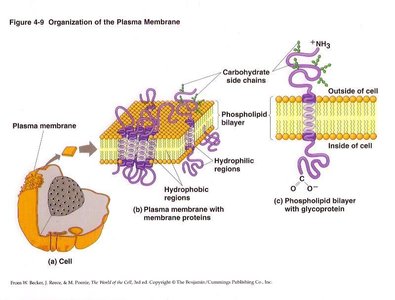
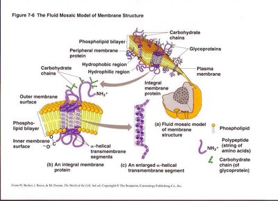
The cell membrane is a dynamic structure essential for maintaining cellular integrity and regulating transport. The understanding of membrane structure has evolved, culminating in the fluid mosaic model, which describes the membrane as a flexible bilayer of lipids with embedded proteins.
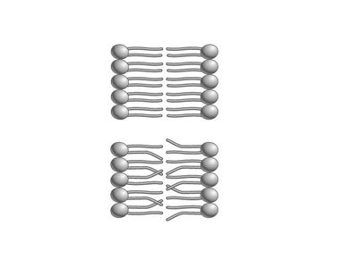
Phospholipid bilayer: Forms the basic structural framework.
Membrane proteins: Integral and peripheral proteins contribute to function and structure.
Carbohydrate side chains: Involved in cell recognition and signaling.
Membrane Fluidity: Saturated vs. Unsaturated Fats
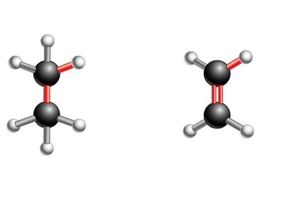
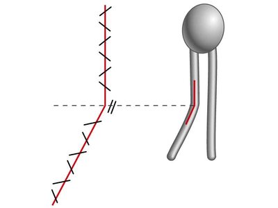
Membrane fluidity is influenced by the chemical structure of its lipid components. Saturated and unsaturated fatty acids affect how tightly the lipid tails pack together, impacting membrane flexibility.
Saturated fatty acids: No double bonds; tails pack closely, making the membrane less fluid.
Unsaturated fatty acids: Contain double bonds; tails are kinked and less tightly packed, increasing fluidity.
Biological relevance: Animal fats (e.g., butter) are saturated and solid at room temperature; plant fats (e.g., corn oil) are unsaturated and fluid.

Classes and Functions of Membrane Lipids
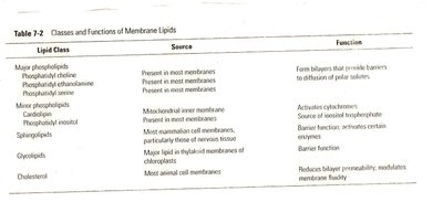
Membranes contain various lipid classes, each with specific sources and functions. These include phospholipids, glycolipids, sphingolipids, and cholesterol.
Phospholipids: Form bilayers that provide barriers to diffusion of polar solutes.
Glycolipids: Barrier function, especially in chloroplasts.
Cholesterol: Reduces bilayer permeability and modulates membrane fluidity.
Lipid Class | Source | Function |
|---|---|---|
Phosphatidylcholine, Phosphatidylethanolamine, Phosphatidylserine | Present in most membranes | Form bilayers; barrier to polar solutes |
Cardiolipin | Mitochondrial inner membrane | Activates cytochrome |
Sphingolipids | Most mammalian cell membranes | Barrier function, structure |
Glycolipids | Major lipid in thylakoid membranes of chloroplasts | Barrier function |
Cholesterol | Most animal cell membranes | Reduces permeability, modulates fluidity |
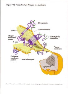
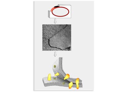
Visualizing Membrane Proteins: Freeze-Fracture and Electron Microscopy
Membrane proteins can be visualized using freeze-fracture techniques and electron microscopy. These methods reveal the distribution and structure of proteins within the lipid bilayer.
Freeze-fracture: Splits the membrane, exposing the interior and revealing pits and mounds caused by proteins.
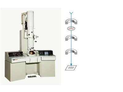
Electron microscopy: Allows visualization of structures smaller than the wavelength of light, such as membrane proteins.
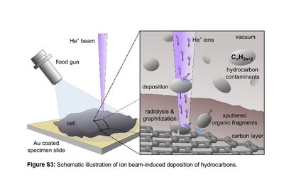
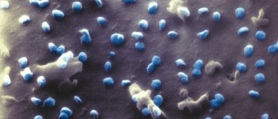
Helium Ion Microscopy (HIM)
Helium ion microscopy is a modern technique for imaging biological samples at high resolution without the need for conductive coatings. It provides pristine images of cell surfaces and membrane structures.
Charge compensation: Allows imaging of insulating samples.
Higher resolution: Reveals fine details of membrane structure.
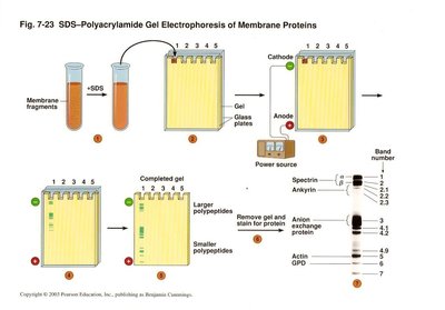
Isolation and Study of Membrane Proteins
Detergents are used to isolate membrane proteins by breaking up the lipid bilayer and coating hydrophobic regions. This enables purification and detailed study of membrane proteins.
Detergents: Form micelles in water, disrupt membranes, and solubilize proteins.
Polyacrylamide Gel Electrophoresis (PAGE): Separates proteins based on size for analysis.
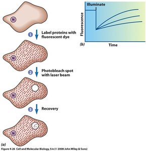
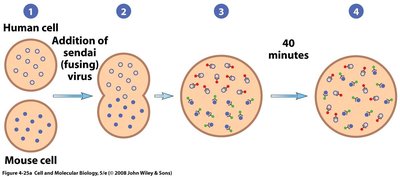
Membrane Fluidity and Mobility: Experimental Demonstrations
The fluidity and mobility of cell membranes can be demonstrated using several experimental techniques, supporting the fluid mosaic model.
Laser bleaching: Fluorescently labeled proteins are photobleached, and recovery indicates protein mobility.
Membrane hybridization: Fusion of cells with different membrane proteins shows mixing and mobility.
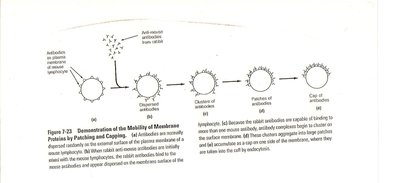
Patching and capping: Antibodies cause clustering of membrane proteins, demonstrating lateral movement.
Additional info:
These notes cover foundational concepts from cell biology chapters including cell types, cell structure, membrane history, membrane chemistry, and experimental techniques for studying membranes. The content is expanded for clarity and completeness, suitable for college-level exam preparation.
