 Back
BackExtracellular Matrix and Cell Junctions in Animal Cells
Study Guide - Smart Notes
 Tailored notes based on your materials, expanded with key definitions, examples, and context.
Extracellular Matrix (ECM) of Animal Cells
Structure and Functions of the ECM
The extracellular matrix (ECM) is a complex network of macromolecules located immediately outside the plasma membrane of animal cells. It provides structural support, organizes tissue architecture, facilitates cell movement, and mediates signaling between cells and their environment.
Support and Organization: The ECM maintains the three-dimensional structure of tissues.
Traction for Cell Movement: The ECM provides a substrate for cell migration, essential during development, wound healing, and immune responses.
Signaling: The ECM transmits signals that regulate cell growth, differentiation, and survival.
The components of the ECM are synthesized and secreted by cells. Major components include:
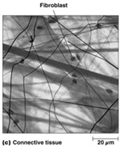
Collagens and Elastins: Structural fibers that provide strength and elasticity.
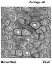
Proteoglycans: Hydrated gel-forming molecules that fill the extracellular space.
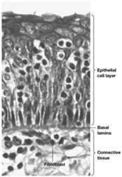
Fibronectins and Laminins: Adhesive glycoproteins that connect cells to the ECM.
Collagens and Elastins: Structural Fibers
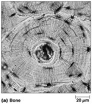
Collagen: The most abundant protein in vertebrates, forming a rigid triple helix that provides tensile strength. Collagen crosslinking increases with age, reducing tissue flexibility. Defects in collagen can lead to diseases such as fibrosis and scarring.
Elastin: Forms crosslinked fibrous networks that can stretch and recoil, imparting elasticity to tissues like skin, lungs, arteries, and intestines. Elastin content decreases with aging.
Proteoglycans: Matrix/Gel Components
Proteoglycans are heavily glycosylated proteins composed of a core protein and one or more glycosaminoglycan (GAG) chains. They are synthesized in the rough ER, glycosylated in the Golgi, and secreted into the ECM.
GAGs: Examples include heparan sulfate and hyaluronic acid.
Proteoglycans provide hydration and resistance to compression.
Fibronectins and Laminins: Adhesive Molecules
Fibronectin: Serves as a bridge connecting cells to the ECM by binding both collagen and integrins.
Laminin: A large glycoprotein found in the basal lamina, important for tissue structure and cell adhesion.
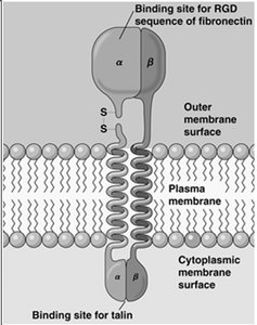
Integrins: ECM Receptors
Integrins are transmembrane receptors that connect the ECM to the cytoskeleton. They are heterodimers composed of α and β subunits. Integrins bind fibronectin and laminin extracellularly and interact with the cytoskeleton intracellularly.
Integrins mediate cell attachment, movement, and signal transduction.
Normal cells require anchorage to the ECM for growth (anchorage-dependent growth), while cancer cells can grow without attachment (anchorage-independent growth).
Cell-Cell Adhesion Proteins
Types and Functions
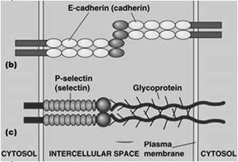
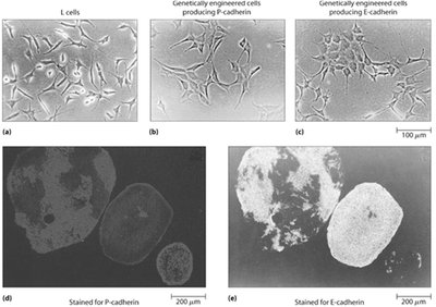
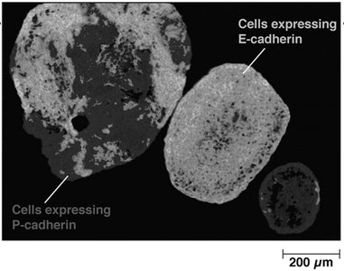
Cell-cell adhesion is mediated by transmembrane proteins (adhesion receptors) on adjacent cells. These interactions can be homophilic (same protein on both cells) or heterophilic (different proteins or ECM components).
Most adhesion receptors are linked to the cytoskeleton.
Some serve as receptors for pathogens.
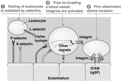
Leukocyte Rolling and Arrest
Leukocyte migration during inflammation involves sequential interactions with endothelial cells:
Selectins: Mediate initial rolling of leukocytes along the endothelium.
Integrins: Become activated and mediate firm adhesion, allowing leukocytes to invade tissues.
ICAM-1: An intercellular adhesion molecule that binds integrins.
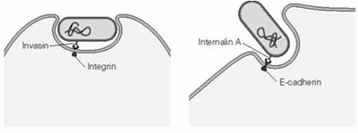
Pathogen Interaction with Adhesion Proteins
Certain pathogens exploit cell adhesion proteins to invade host cells:
Yersinia pestis: Uses integrins for entry.
Listeria monocytogenes: Uses E-cadherin for entry.
Human rhinovirus (HRV-14): Uses ICAM-1 as a receptor.
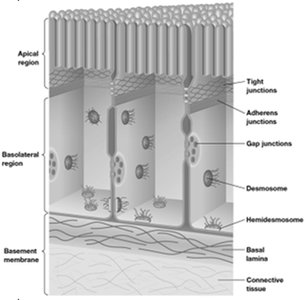
Cell Junctions in Animal Cells
Types of Cell Junctions
Animal cells are connected by specialized junctions that serve different functions:
Adhesive Junctions: Mediate cell-to-cell or cell-to-ECM connections, often involving cadherins or integrins.
Tight Junctions: Form permeability barriers and maintain cell polarity.
Gap Junctions: Allow direct cytoplasmic communication between adjacent cells.
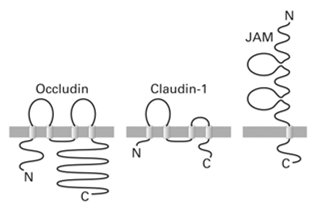
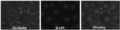
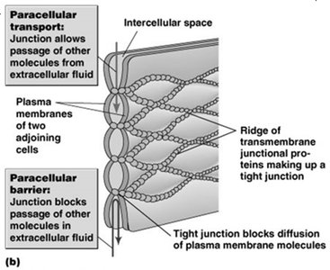
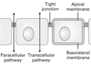
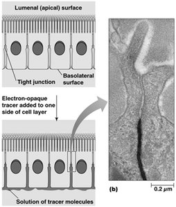
Tight Junctions
Tight junctions are composed of proteins such as claudin, occludin, and JAM. They seal the space between adjacent cells, preventing the passage of molecules and maintaining distinct apical and basolateral membrane domains.
Establish cell polarity by preventing lateral diffusion of membrane proteins and lipids.
Control paracellular transport (movement of substances between cells).

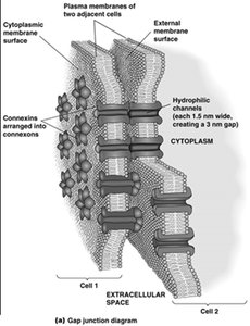
Gap Junctions
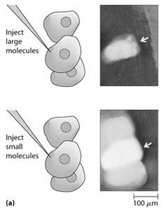
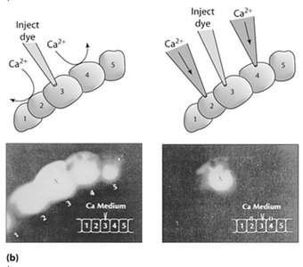
Gap junctions are clusters of intercellular channels that allow the passage of ions and small molecules between adjacent cells. Each channel is formed by the alignment of two connexons (hemichannels), each composed of six connexin proteins.
Enable electrical and metabolic coupling between cells.
Small molecules (e.g., Ca2+, cAMP) can pass through; elevated Ca2+ closes the junction to prevent cell death from spreading.

Comparison: Plasmodesmata in Plants
Plasmodesmata are cytoplasmic channels in plant cell walls that allow direct communication between adjacent cells. They are functionally analogous to gap junctions in animal cells.
Permit the movement of ions, small molecules, and some proteins and RNA between plant cells.
