 Back
BackMeiosis, Sexual Reproduction, and Genetic Variation: Study Guide
Study Guide - Smart Notes
 Tailored notes based on your materials, expanded with key definitions, examples, and context.
Meiosis and Sexual Reproduction
Overview of Reproduction Types
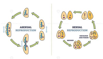
Reproduction in eukaryotes occurs via two main mechanisms: asexual reproduction and sexual reproduction. Each process has distinct cellular and genetic outcomes.
Asexual reproduction: Involves cell division by mitosis, producing genetically identical diploid cells (2n → 2n).
Sexual reproduction: Combines genetic material from two parents, involving meiosis to produce haploid gametes (1n), which fuse to form a genetically unique diploid organism.
Genetic variety: Sexual reproduction introduces new combinations of alleles, increasing diversity among offspring.
Mitosis: Asexual Cell Division
Stages of Mitosis
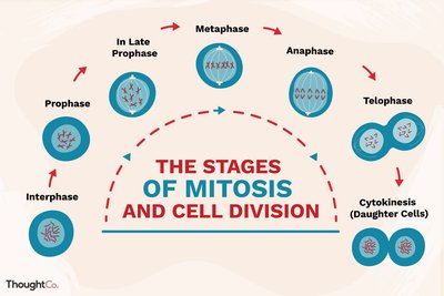
Mitosis is the process by which a cell divides to produce two genetically identical daughter cells. It is essential for growth, repair, and asexual reproduction.
Interphase: Cell prepares for division by replicating DNA and organelles.
Prophase: Chromosomes condense, spindle fibers form.
Late Prophase (Prometaphase): Nuclear envelope breaks down, spindle fibers attach to chromosomes.
Metaphase: Chromosomes align at the cell's equator.
Anaphase: Sister chromatids separate and move to opposite poles.
Telophase: Nuclear envelopes reform, chromosomes decondense.
Cytokinesis: Cytoplasm divides, forming two daughter cells.
Genetic Concepts: Alleles, Genotype, and Phenotype
Definitions and Examples
Genetic variation is fundamental to sexual reproduction. Key terms include:
Gene: A unit of DNA encoding a specific trait.
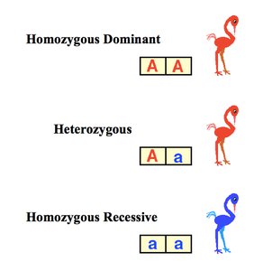
Allele: Different versions of a gene (e.g., blue/brown eyes).
Genotype: The genetic makeup of an organism (the alleles present).
Phenotype: Observable traits, influenced by genotype and environment.
Locus: The specific location of a gene on a chromosome.
Homozygous: Both alleles at a locus are the same (AA or aa).
Heterozygous: Two different alleles at a locus (Aa).
Gametes and Life Cycles
Formation and Role of Gametes
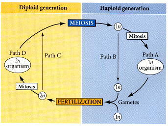
Gametes are specialized haploid cells produced by meiosis for sexual reproduction. Their fusion restores diploidy in the zygote.
Animals: Meiosis produces sperm and ova (haploid gametes).
Plants: Meiosis produces haploid spores, which develop into haploid organisms that produce gametes by mitosis.
Fertilization: Fusion of two haploid gametes forms a diploid zygote.
Meiosis: Process and Stages
Meiosis Overview
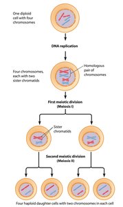
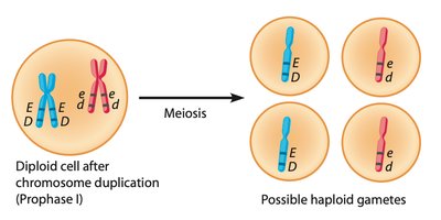
Meiosis is a specialized cell division that reduces chromosome number by half, producing four genetically distinct haploid cells from one diploid cell. It consists of one round of DNA replication followed by two nuclear divisions.
Meiosis I: Homologous chromosomes separate, chromosome number is halved.
Meiosis II: Sister chromatids separate, similar to mitosis.
Cell Cycle and Meiosis
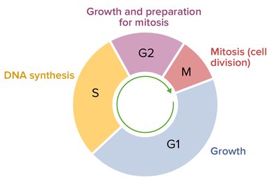
The cell cycle includes phases of growth (G1), DNA synthesis (S), preparation (G2), and division (M). Meiosis occurs during the M phase.
G1: Cell growth.
S: DNA replication.
G2: Preparation for division.
M: Meiosis and cytokinesis.
Meiosis I: Reduction Division
Prophase I: Five Stages
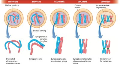
Prophase I is complex and includes five distinct stages, each with specific chromosomal events.
Leptotene: Chromatin condenses, chromosomes become visible.
Zygotene: Homologous chromosomes pair (synapsis), forming tetrads.
Pachytene: Crossing over occurs, exchanging genetic material between homologs.
Diplotene: Tetrads begin to separate, chiasmata (crossing over sites) remain visible.
Diakinesis: Chromosomes fully condense, spindle forms, nuclear envelope breaks down.
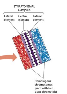
Synapsis and Crossing Over
Synapsis is the pairing of homologous chromosomes, facilitated by the synaptonemal complex. Crossing over exchanges genetic material, increasing genetic diversity.
Synaptonemal complex: Protein structure holding homologs together.
Chiasma: Physical site of crossing over.
Genetic exchange: Occurs between inner chromatids of tetrads.
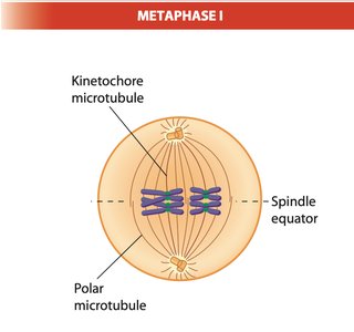
Metaphase I
Tetrads align at the cell equator, attached to spindle fibers. Homologs are held together by chiasmata.
Random alignment: Maternal and paternal homologs are distributed randomly, contributing to genetic variation.

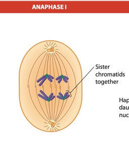
Anaphase I
Homologous chromosomes separate and move to opposite poles. Each daughter cell receives a unique combination of chromosomes.
Source of variation: Division of maternal and paternal chromosomes, plus crossing over.
Telophase I and Cytokinesis
Chromosomes reach the poles, nuclear envelopes may reform, and the cell divides. Two haploid daughter cells are produced, each with "X" shaped chromosomes.
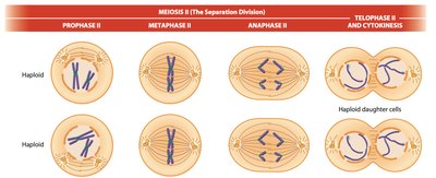
Meiosis II: Separation Division
Overview and Stages
Meiosis II resembles mitosis, separating sister chromatids without further DNA replication. It produces four haploid cells.
Prophase II: Chromosomes condense, spindle forms.
Metaphase II: Chromosomes align at the equator.
Anaphase II: Sister chromatids separate.
Telophase II and Cytokinesis: New nuclei form, cytoplasm divides.
Genetic Recombination and Errors
Crossing Over and Genetic Diversity
Crossing over during prophase I is a major source of genetic diversity. It involves the exchange of DNA segments between homologous chromosomes.
Mechanism: Double helix breakage, strand invasion, and DNA synthesis.
Result: New allele combinations in gametes.

Errors in Crossing Over
Imprecise pairing can lead to unequal crossing over, causing deletions or duplications of DNA segments.
Unequal crossing over: Chiasma forms at incorrect locations.
Consequences: One chromosome may lose DNA (deletion), another may gain extra DNA (duplication).
Nondisjunction and Aneuploidy
Nondisjunction is the failure of homologs or sister chromatids to separate properly during anaphase I or II, resulting in cells with abnormal chromosome numbers.
Aneuploidy: Cells have too many or too few chromosomes.
Monosomy: Missing one chromosome (2n−1).
Trisomy: Extra chromosome (2n+1), e.g., Trisomy 21 (Down syndrome).
Gamete Production in Animals
Sperm and Egg Formation
Sperm and egg production differ in their mechanisms and timing.
Sperm: Four spermatids are produced by meiosis, which differentiate into sperm. Sperm are generated continuously throughout the male's lifetime.
Eggs: Oocytes begin meiosis but pause in diplotene for years. Hormones stimulate completion. Only one haploid cell is produced; three polar bodies degenerate.
Special Cases: Parthenogenesis
Parthenogenesis
Parthenogenesis is a form of reproduction where an egg develops into an offspring without fertilization by sperm. This occurs in some species, such as the Komodo dragon.
Mechanism: Haploid egg fuses with a haploid polar body.
Genetic outcome: Offspring inherit only maternal genes, but are not exact duplicates due to allele combinations.
Comparison: Mitosis vs. Meiosis
Key Differences
Mitosis and meiosis are both forms of cell division, but they serve different purposes and produce different outcomes.
Feature | Mitosis | Meiosis |
|---|---|---|
Number of divisions | One | Two |
Number of daughter cells | Two | Four |
Genetic identity | Identical to parent | Genetically unique |
Chromosome number | Diploid (2n) | Haploid (1n) |
Function | Growth, repair, asexual reproduction | Sexual reproduction |
Summary Table: Stages of Meiosis
Stage | Key Events |
|---|---|
Prophase I | Chromosome condensation, synapsis, crossing over |
Metaphase I | Tetrads align at equator |
Anaphase I | Homologs separate |
Telophase I | Two haploid cells form |
Prophase II | Chromosomes condense, spindle forms |
Metaphase II | Chromosomes align at equator |
Anaphase II | Sister chromatids separate |
Telophase II | Four haploid cells form |
Key Equations
Chromosome number after meiosis:
Genetic combinations: Number of possible gametes = (where n = number of chromosome pairs)
