DNase I cuts DNA that is not protected by bound proteins but is unable to cut DNA that is complexed with proteins. Human DNA is isolated, stripped of its nonhistone proteins, and mixed with DNase I. Samples are removed after 30 minutes, 1 hour, and 4 hours and run separately in gel electrophoresis. The resulting gel is stained to make all DNA fragments in it visible, and the results are shown in the figure. DNA fragment sizes in base pairs (bp) are estimated by the scale to the left of the gel. Examine the gel results and speculate why longer DNase I treatment produces different results.
Table of contents
- 1. Introduction to Genetics51m
- 2. Mendel's Laws of Inheritance3h 37m
- 3. Extensions to Mendelian Inheritance2h 41m
- 4. Genetic Mapping and Linkage2h 28m
- 5. Genetics of Bacteria and Viruses1h 21m
- 6. Chromosomal Variation1h 48m
- 7. DNA and Chromosome Structure56m
- 8. DNA Replication1h 10m
- 9. Mitosis and Meiosis1h 34m
- 10. Transcription1h 0m
- 11. Translation58m
- 12. Gene Regulation in Prokaryotes1h 19m
- 13. Gene Regulation in Eukaryotes44m
- 14. Genetic Control of Development44m
- 15. Genomes and Genomics1h 50m
- 16. Transposable Elements47m
- 17. Mutation, Repair, and Recombination1h 6m
- 18. Molecular Genetic Tools19m
- 19. Cancer Genetics29m
- 20. Quantitative Genetics1h 26m
- 21. Population Genetics50m
- 22. Evolutionary Genetics29m
7. DNA and Chromosome Structure
Eukaryotic Chromosome Structure
Problem 30
Textbook Question
Because of its rapid turnaround time, fluorescent in situ hybridization (FISH) is commonly used in hospitals and laboratories as an aneuploid screen of cells retrieved from amniocentesis and chorionic villus sampling (CVS). Chromosomes 13, 18, 21, X, and Y are typically screened for aneuploidy in this way. Explain how FISH might be accomplished using amniotic or CVS samples and why the above chromosomes have been chosen for screening.
 Verified step by step guidance
1
Step 1: Begin by explaining the concept of fluorescent in situ hybridization (FISH). FISH is a molecular cytogenetic technique that uses fluorescently labeled DNA probes to bind to specific chromosome regions. This allows visualization of specific genetic sequences under a fluorescence microscope.
Step 2: Describe the process of sample preparation. Cells from amniotic fluid or chorionic villus sampling (CVS) are collected and cultured. The chromosomes within these cells are then fixed onto a slide to prepare them for hybridization with the fluorescent probes.
Step 3: Explain the hybridization process. Fluorescently labeled DNA probes complementary to sequences on chromosomes 13, 18, 21, X, and Y are applied to the slide. These probes bind specifically to their target sequences on the chromosomes, allowing visualization of these regions.
Step 4: Discuss the detection and analysis. After hybridization, the slide is examined under a fluorescence microscope. The presence, absence, or abnormal number of fluorescent signals indicates whether aneuploidy (abnormal chromosome number) is present for the screened chromosomes.
Step 5: Explain the rationale for choosing chromosomes 13, 18, 21, X, and Y. These chromosomes are associated with common aneuploid conditions, such as trisomy 13 (Patau syndrome), trisomy 18 (Edwards syndrome), trisomy 21 (Down syndrome), and sex chromosome abnormalities (e.g., Turner syndrome and Klinefelter syndrome). Screening these chromosomes provides critical information about the most clinically relevant aneuploidies.
 Verified video answer for a similar problem:
This video solution was recommended by our tutors as helpful for the problem above
Video duration:
2mPlay a video:
Was this helpful?
Key Concepts
Here are the essential concepts you must grasp in order to answer the question correctly.
Fluorescent In Situ Hybridization (FISH)
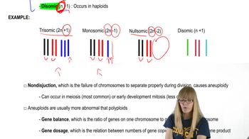
FISH is a molecular cytogenetic technique that uses fluorescent probes to bind to specific chromosome regions, allowing visualization of chromosomal abnormalities. In the context of amniotic fluid or CVS samples, FISH can quickly identify the presence or absence of specific chromosomes, making it a valuable tool for detecting aneuploidies, such as trisomy or monosomy.
Recommended video:
Guided course

Functional Genomics
Aneuploidy
Aneuploidy refers to an abnormal number of chromosomes in a cell, which can lead to genetic disorders. Common aneuploidies include trisomy 21 (Down syndrome), trisomy 18 (Edwards syndrome), and trisomy 13 (Patau syndrome). Screening for these specific chromosomes during prenatal testing is crucial because they are associated with significant developmental and health issues.
Recommended video:
Guided course

Aneuploidy
Amniocentesis and Chorionic Villus Sampling (CVS)
Amniocentesis and CVS are prenatal diagnostic procedures used to obtain fetal cells for genetic analysis. Amniocentesis involves extracting amniotic fluid, while CVS involves sampling placental tissue. Both methods provide valuable samples for FISH analysis, enabling early detection of chromosomal abnormalities in the fetus.
Recommended video:
Guided course

Trihybrid Cross
Related Videos
Related Practice
Textbook Question
360
views


