Following the tragic shooting of 20 children at a school in Newtown, Connecticut, in 2012, Connecticut's state medical examiner requested a full genetic analysis of the killer's genome. What do you think investigators might be looking for? What might they expect to find? Might this analysis lead to an oversimplified analysis of the cause of the tragedy?
Table of contents
- 1. Introduction to Genetics51m
- 2. Mendel's Laws of Inheritance3h 37m
- 3. Extensions to Mendelian Inheritance2h 41m
- 4. Genetic Mapping and Linkage2h 28m
- 5. Genetics of Bacteria and Viruses1h 21m
- 6. Chromosomal Variation1h 48m
- 7. DNA and Chromosome Structure56m
- 8. DNA Replication1h 10m
- 9. Mitosis and Meiosis1h 34m
- 10. Transcription1h 0m
- 11. Translation58m
- 12. Gene Regulation in Prokaryotes1h 19m
- 13. Gene Regulation in Eukaryotes44m
- 14. Genetic Control of Development44m
- 15. Genomes and Genomics1h 50m
- 16. Transposable Elements47m
- 17. Mutation, Repair, and Recombination1h 6m
- 18. Molecular Genetic Tools19m
- 19. Cancer Genetics29m
- 20. Quantitative Genetics1h 26m
- 21. Population Genetics50m
- 22. Evolutionary Genetics29m
18. Molecular Genetic Tools
Methods for Analyzing DNA
Problem 27b
Textbook Question
Genomic DNA from the nematode worm Caenorhabditis elegans is organized by nucleosomes in the manner typical of eukaryotic genomes, with 145 bp encircling each nucleosome and approximately 55 bp in linker DNA. When C. elegans chromatin is carefully isolated, stripped of nonhistone proteins, and placed in an appropriate buffer, the chromatin decondenses to the 10-nm fiber structure. Suppose researchers mix a sample of 10-nm–fiber chromatin with a large amount of the enzyme DNase I that randomly cleaves DNA in regions not protected by bound protein. Next, they remove the nucleosomes, separate the DNA fragments by gel electrophoresis, and stain all the DNA fragments in the gel.
Explain the origin of DNA fragments seen in the gel.
 Verified step by step guidance
1
Step 1: Understand the structure of chromatin in Caenorhabditis elegans. Chromatin consists of nucleosomes, which are DNA segments of 145 base pairs (bp) wrapped around histone proteins, and linker DNA, which is approximately 55 bp long and connects adjacent nucleosomes. The nucleosomes protect the DNA they encircle from enzymatic cleavage.
Step 2: Recognize the role of DNase I in the experiment. DNase I is an enzyme that randomly cleaves DNA in regions not protected by bound proteins, such as the linker DNA between nucleosomes. The DNA wrapped around histones is shielded from DNase I activity.
Step 3: Consider the experimental setup. After treating the chromatin with DNase I, the researchers remove the nucleosomes, leaving behind DNA fragments that were protected by histones and fragments that were cleaved in the linker regions.
Step 4: Analyze the DNA fragments using gel electrophoresis. Gel electrophoresis separates DNA fragments based on their size. The protected DNA fragments (145 bp) and cleaved linker DNA fragments (approximately 55 bp or smaller) will appear as distinct bands on the gel.
Step 5: Explain the origin of the DNA fragments seen in the gel. The bands correspond to DNA fragments protected by nucleosomes (145 bp) and smaller fragments resulting from DNase I cleavage of linker DNA. The pattern reflects the periodic organization of chromatin into nucleosomes and linker regions.
 Verified video answer for a similar problem:
This video solution was recommended by our tutors as helpful for the problem above
Video duration:
2mPlay a video:
Was this helpful?
Key Concepts
Here are the essential concepts you must grasp in order to answer the question correctly.
Nucleosome Structure
Nucleosomes are the fundamental units of chromatin, consisting of DNA wrapped around a core of histone proteins. Each nucleosome typically contains about 145 base pairs of DNA, which is protected from enzymatic cleavage. Understanding nucleosome structure is crucial for explaining how certain regions of DNA remain intact during experiments involving DNase I, as the nucleosomes shield the DNA from being cut.
Recommended video:
Guided course

Chromosome Structure
DNase I Activity
DNase I is an enzyme that cleaves DNA at sites that are not protected by proteins, such as histones in nucleosomes. When chromatin is treated with DNase I, the enzyme will cut the linker DNA and any exposed regions of DNA, leading to the generation of fragments. This concept is essential for understanding the resulting DNA fragments observed in gel electrophoresis after nucleosome removal.
Recommended video:
Guided course

Decision Between Lytic and Lysogenic Cycles
Gel Electrophoresis
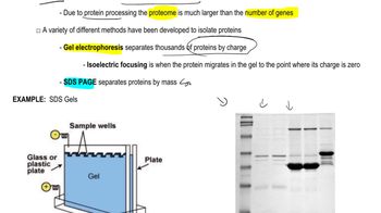
Gel electrophoresis is a laboratory technique used to separate DNA fragments based on their size. When DNA fragments are subjected to an electric field in a gel matrix, smaller fragments migrate faster than larger ones. This method allows researchers to visualize the DNA fragments generated by DNase I cleavage, providing insights into the organization and accessibility of chromatin in C. elegans.
Recommended video:
Guided course

Proteomics

 7:40m
7:40mWatch next
Master Methods for Analyzing DNA and RNA with a bite sized video explanation from Kylia
Start learningRelated Videos
Related Practice
Textbook Question
344
views
