The flow of genetic information from DNA to protein is mediated by messenger RNA. If you introduce short DNA strands (called antisense oligonucleotides) that are complementary to mRNAs, hydrogen bonding may occur and 'label' the DNA/RNA hybrid for ribonuclease-H degradation of the RNA. One study [Lloyd et al. (2001). Nucl. Acids Res. 29:3664–3673] compared the effect of different-length antisense oligonucleotides upon ribonuclease-H–mediated degradation of tumor necrosis factor (TNFα) mRNA. TNFα exhibits antitumor and pro-inflammatory activities. The following graph indicates the efficacy of various-sized antisense oligonucleotides in causing ribonuclease-H cleavage. Describe how antisense oligonucleotides interrupt the flow of genetic information in a cell.
11. Translation
Translation
Problem 39l
Textbook Question
Answer the following questions about the accompanying diagram.
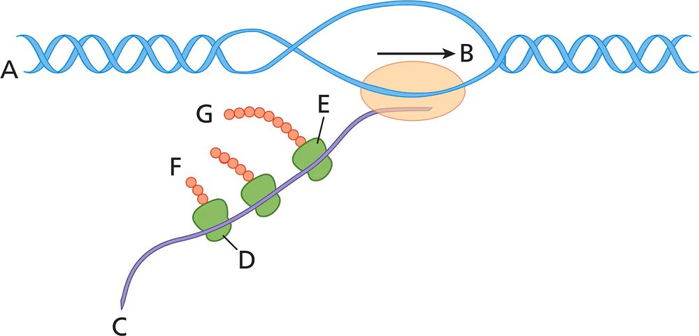
Does the diagram depict molecular activity in a bacterium or a eukaryote? Explain the reasoning for your answer.
 Verified step by step guidance
1
Examine the diagram carefully. The image depicts a process involving DNA (labeled A), RNA (labeled C), and ribosomes (labeled D, E, F) synthesizing polypeptides (labeled G). This suggests the process of transcription and translation.
Identify the key features of the diagram. The RNA strand (C) is being synthesized from the DNA template (A) by the enzyme RNA polymerase (labeled B). Ribosomes (D, E, F) are attached to the RNA strand, actively translating it into polypeptides (G).
Consider the spatial arrangement of transcription and translation. In bacteria, transcription and translation occur simultaneously in the cytoplasm because there is no nuclear membrane separating these processes. In eukaryotes, transcription occurs in the nucleus, and translation occurs in the cytoplasm after RNA processing.
Note the absence of RNA processing steps such as splicing, capping, or polyadenylation in the diagram. These steps are characteristic of eukaryotic cells but are absent in bacterial cells.
Conclude based on the evidence. The simultaneous transcription and translation depicted in the diagram, along with the absence of RNA processing, strongly suggests that the molecular activity is occurring in a bacterium rather than a eukaryote.
Verified video answer for a similar problem:This video solution was recommended by our tutors as helpful for the problem above
Video duration:
2mWas this helpful?
Key Concepts
Here are the essential concepts you must grasp in order to answer the question correctly.
Translation Process
Translation is the process by which ribosomes synthesize proteins using messenger RNA (mRNA) as a template. During translation, ribosomes read the sequence of codons in mRNA and assemble the corresponding amino acids into a polypeptide chain. This process is crucial for gene expression and occurs in both prokaryotic and eukaryotic cells, although the mechanisms and locations differ.
Recommended video:
Guided course

 08:39
08:39mRNA Processing
Prokaryotic vs. Eukaryotic Cells
Prokaryotic cells, such as bacteria, lack a defined nucleus and membrane-bound organelles, while eukaryotic cells have a nucleus and various organelles. In prokaryotes, translation occurs in the cytoplasm simultaneously with transcription, whereas in eukaryotes, transcription occurs in the nucleus and translation occurs in the cytoplasm. This distinction is essential for determining the cellular context depicted in the diagram.
Recommended video:
Guided course
 10:14
10:14Prokaryotic Transcription
Ribosomes and Their Structure
Ribosomes are complex molecular machines made of ribosomal RNA (rRNA) and proteins that facilitate the translation of mRNA into proteins. They consist of two subunits (large and small) that come together during translation. The presence and structure of ribosomes can indicate whether the diagram represents a prokaryotic or eukaryotic cell, as eukaryotic ribosomes are larger and more complex than those found in prokaryotes.
Recommended video:
Guided course
 03:53
03:53Ribosome Structure
 7:58m
7:58mWatch next
Master Translation initiation with a bite sized video explanation from Kylia
Start learningRelated Videos
Related Practice
Textbook Question
1082
views