 Back
BackAvian In Ovo Assay for Sex Steroid Hormone Disruption: Genetics and Morphological Analysis in Japanese Quail
Study Guide - Smart Notes
 Tailored notes based on your materials, expanded with key definitions, examples, and context.
Avian In Ovo Assay for Sex Steroid Hormone Disrupting Properties
Introduction and Purpose
This protocol describes an avian in ovo assay using Japanese quail embryos to evaluate the estrogenic and anti-estrogenic effects of chemical substances. The assay focuses on the disruption of sex steroid hormone action during embryonic development, with particular attention to the genetic and morphological differentiation of reproductive organs. The method integrates both genetic sex determination and detailed morphological analysis, making it highly relevant to studies in genetics, developmental biology, and endocrine disruption.
Background: Sex Determination and Differentiation in Birds
Sexual Differentiation in Avian Species
Female birds (ZW) develop an ovary and oviduct (Müllerian duct, MD) on the left side; the right ovary and MD regress under estrogen influence.
Male birds (ZZ) develop bilateral testes; MDs regress due to minimal estrogen synthesis during this period.
Estrogen is critical for sex-specific development of reproductive organs. Disruption by exogenous chemicals can cause abnormal organ morphology.
Genetic sex determination is essential, as morphological sex can be altered by hormonal disruption.
Example: Exposure to estrogenic compounds (e.g., 17α-ethinylestradiol) can cause residual MDs and ovotestis formation in males, and hypertrophy or persistence of the right MD in females.
Assay Overview and Workflow
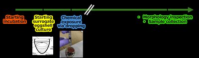
Experimental Timeline and Key Steps
Incubation of fertilized Japanese quail eggs (E0).
Transfer of embryo to surrogate eggshell (E2.5).
Chemical exposure via dropping test solution onto egg contents (E4.5).
Dissection and sample collection for morphological and genetic analysis (E11.5).
Technical Equipment and Materials
Essential Tools
Egg incubator with humidity and temperature control
Eggshell cutter
Syringes, needles, or pipettes for chemical administration
Dissection tools
Stereomicroscope with camera
Image analysis software (e.g., ImageJ)
PCR equipment for genetic sex determination
Preparation of Test Items and Controls
Chemicals and Vehicles
Test solutions stored in glass vials; details such as CAS number, purity, and source must be recorded.
Vehicles: Sesame oil or sodium carboxymethyl cellulose (CMC-Na) preferred; PBS or ddH2O if soluble; DMSO/EtOH as auxiliary agents if justified.
Vehicle control (VC) and positive control (PC, e.g., 17α-ethinylestradiol) are mandatory for assay validation.
Assay Process
Egg Handling and Surrogate Eggshell Culture
Fertilized eggs are selected based on weight and stored at 12–15°C, 60–70% humidity, used within one week.
Incubation at 37.5 ± 0.5°C, 60–70% humidity, with automatic turning.
Surrogate eggshells are prepared from large eggs; embryos are transferred at E2.5 and sealed with cling wrap.
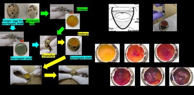
Chemical Exposure and Embryo Viability
Test solution (50 µl) is administered at E4.5 by dropping onto the egg contents through the surrogate shell.
Embryo survival is monitored daily until E11.5; survival rate is calculated based on initial numbers at exposure.
Dissection and Sample Collection
Blood is collected for genetic sex determination by PCR.
Embryos are dissected, and internal reproductive organs are photographed for morphological analysis.
Phenotypic sex is recorded, but final sex assignment is based on genetic analysis.
Genetic Sex Determination by PCR
Principle and Method
Avian sex chromosomes: Z and W; CHD1Z (Z-linked) and CHD1W (W-linked) genes are targeted.
PCR with specific primers distinguishes males (one band, ZZ) from females (two bands, ZW) on agarose gel.
Example: Direct PCR from blood using PlatinumTM Direct PCR Universal Master Mix; see table below for primer sequences and cycling conditions.
Gene | Forward Primer (2550F) | Reverse Primer (2718R) | Product Size (bp) |
|---|---|---|---|
CHD1W | GTTACTGATTCGTCTACGAGA | ATTGAAATGATCCAGTGCTTG | 450 |
CHD1Z | GTTACTGATTCGTCTACGAGA | ATTGAAATGATCCAGTGCTTG | 600 |
Step | Cycles | Temperature | Time |
|---|---|---|---|
Activation | 1 | 94°C | 2 min |
Denaturation | 40 | 94°C | 15 sec |
Annealing | 40 | 60°C | 15 sec |
Extension | 40 | 68°C | 20 sec |
Final Extension | 1 | 68°C | 5 min |
Hold | 1 | 4°C | Hold |

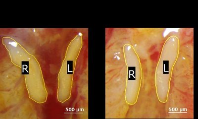
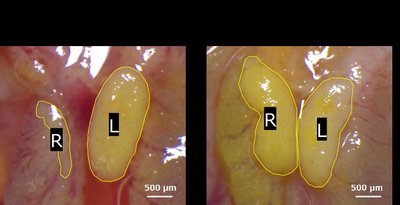
Morphological Assessment of Reproductive Organs
Male Embryos: Müllerian Ducts and Testes
Presence and area of left/right MDs are measured; area is normalized by $\text{BW}^{2/3}$ to correct for body size.
Abnormalities (cysts, edema) are recorded and compared across groups.
Testis size (left and right) and their ratio are measured and compared.
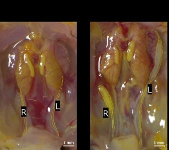
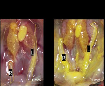
Female Embryos: Müllerian Ducts and Ovaries
Area of left and right MDs is measured and normalized by $\text{BW}^{2/3}$.
Ovary size (left and right) is measured; right ovary regression is a normal feature, and persistence indicates disruption.
Data Interpretation and Statistical Analysis
Criteria for Estrogenic and Anti-Estrogenic Effects
Estrogenic effects: Increased MD area in males, decreased right testis size, increased left/right testis ratio, increased right MD area in females.
Anti-estrogenic/aromatase-inhibiting effects: Increased right ovary area in females, decreased left ovary area.
Statistical analysis typically involves two-way repeated measures ANOVA for area comparisons and Fisher’s exact test for abnormality rates. Alternative methods are used if data do not meet assumptions.
Validity Control and General Toxicity
Ensuring Experimental Reliability
Positive controls must show significant differences from vehicle controls for assay validity.
Sample size: At least 6 males and 6 females per group for statistical power.
General toxicity is assessed by comparing body weight and developmental stage; significant reductions (except at highest dose) indicate non-specific toxicity.
Summary Table: Key Indicators and Interpretation
Indicator | Interpretation |
|---|---|
Increased MD area in males | Estrogenic effect |
Decreased right testis size in males | Estrogenic effect |
Increased left/right testis ratio in males | Estrogenic effect |
Increased right MD area in females | Estrogenic effect |
Increased right ovary area in females | Anti-estrogenic/aromatase-inhibiting effect |
Decreased left ovary area in females | Estrogen disrupting effect |
References
Key references are provided for further reading on avian sex differentiation, PCR-based sexing, and the effects of endocrine disruptors on avian development.
Additional info: This protocol is directly relevant to genetics (Ch. 3, 4, 7, 11, 12, 13, 14, 18) as it integrates molecular sex determination, gene-environment interactions, and developmental genetics in a model organism.
