 Back
BackCell Division and Chromosome Heredity: Structured Study Notes
Study Guide - Smart Notes
 Tailored notes based on your materials, expanded with key definitions, examples, and context.
Cell Division and Chromosome Heredity
Introduction to Cell Division
Cell division is a fundamental process in multicellular organisms, enabling growth, development, and maintenance. Accurate transmission of genetic information is essential for organismal integrity and heredity.
Cell division occurs continuously in multicellular organisms, replacing aged or dead cells.
Mother cells transmit genetic information to daughter cells, requiring precise mechanisms for genetic fidelity.
Millions of cells divide every minute in adult humans, especially in tissues like blood.
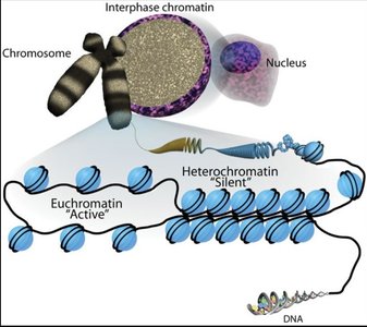
Chromatin and Chromosome Structure
Chromatin is the complex of DNA, RNA, and proteins that forms chromosomes. Chromatin exists in two main forms, euchromatin and heterochromatin, which differ in compaction and gene activity.
Chromatin: Material composing eukaryotic chromosomes.
Euchromatin: Less compact, gene-rich, transcriptionally active.
Heterochromatin: Densely packed, gene-poor, transcriptionally silent.
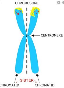
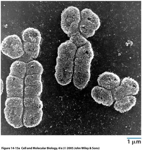
Chromosome: DNA-containing structure with a centromere; consists of two sister chromatids after S-phase.
Homologous chromosomes: Chromosomes with the same structural features and gene patterns.
Chromatid: One of the replicated structures; sister chromatids are the two identical copies joined at the centromere.
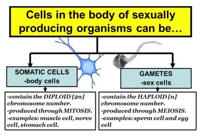
Cell Types and Ploidy
Cells in sexually reproducing organisms are classified as somatic or gametic, differing in chromosome number and function.
Diploid (2n): Two sets of chromosomes, typical of somatic cells.
Haploid (1n): One set of chromosomes, typical of gametes.
Somatic cells: Non-reproductive, usually diploid.
Gametes: Reproductive cells, haploid, produced by meiosis.
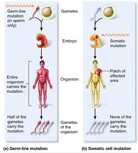
Germ Line vs Somatic Mutations
Mutations can occur in germ-line or somatic cells, affecting inheritance and organismal phenotype differently.
Germ-line mutations: Affect gametes, can be passed to offspring.
Somatic mutations: Affect body cells, not inherited, may cause localized effects.
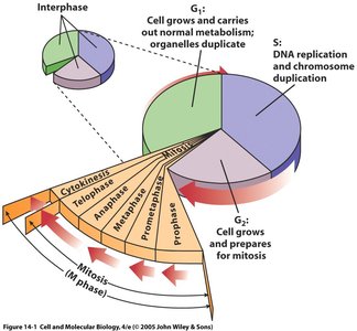
The Cell Cycle
Phases of the Cell Cycle
The cell cycle consists of interphase and M phase, each with distinct roles in cell growth and division.
Interphase: Includes G1 (growth), S (DNA synthesis), and G2 (preparation for mitosis).
M phase: Includes mitosis (nuclear division) and cytokinesis (cytoplasmic division).
Most cells spend the majority of their time in interphase, performing normal functions and preparing for division.
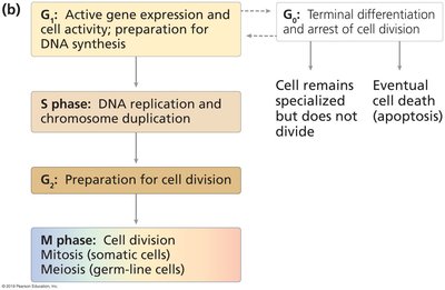
Cell Cycle Regulation
Cell cycle progression is tightly regulated to ensure proper division and prevent errors.
G1: Active gene expression and cell activity.
S: DNA replication and chromosome duplication.
G2: Preparation for cell division.
M: Cell division (mitosis or meiosis).
G0: Terminal differentiation and arrest of cell division.
Mitosis: Division of Somatic Cells
Overview of Mitosis
Mitosis is the process by which somatic cells divide, producing two genetically identical daughter cells. It maintains chromosome number and is essential for growth, maintenance, and repair.
Occurs in both diploid and haploid cells.
Divided into five stages: prophase, prometaphase, metaphase, anaphase, telophase.
Stages of Mitosis
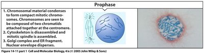
Prophase
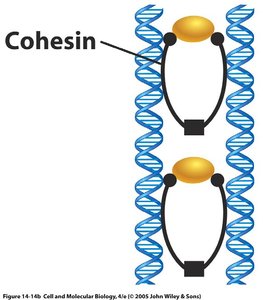
Chromosomes condense, mitotic machinery assembles, and sister chromatids are held together by cohesin.
Chromatin condenses into visible chromosomes.
Sister chromatids are joined by cohesin complexes.

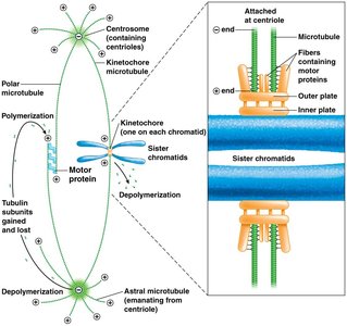
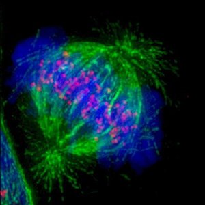
Spindle Formation
Microtubules organize into spindle fibers, facilitating chromosome movement.
Kinetochore microtubules: Attach to chromosomes and direct movement.
Polar microtubules: Extend between centrosomes.
Astral microtubules: Anchor to cell membrane.
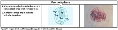
Prometaphase
Nuclear envelope dissolves, spindle assembly completes, and chromosomes move to the cell center.
Microtubules attach to kinetochores.
Chromosomes are positioned at the spindle equator.
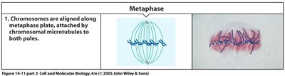
Metaphase
Chromosomes align at the metaphase plate, each chromatid attached to spindle fibers from opposite poles.
Ensures equal segregation of genetic material.
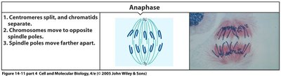
Anaphase
Sister chromatids separate and move toward opposite poles, driven by microtubule shortening.
Centromeres split, chromatids become individual chromosomes.
Spindle poles move farther apart.

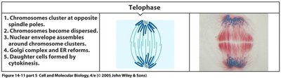
Telophase
Chromosomes reach poles, nuclear envelope reforms, and chromosomes decondense.
Cells return to interphase condition.
Chromosomes disperse and become less visible.

Cytokinesis
Cytokinesis divides the cell into two daughter cells, usually coordinated with mitosis.
Begins in late anaphase with cell surface indentation.
Indentation deepens to form a furrow, encircling the cell.
Mitosis Summary
Start: One diploid cell.
End: Two diploid cells, genetically identical.
Meiosis: Division for Sexual Reproduction
Overview of Meiosis
Meiosis produces haploid gametes, enabling sexual reproduction and genetic diversity. It involves two divisions (meiosis I and II) and recombination.
Meiosis I: Separation of homologous chromosomes.
Meiosis II: Separation of sister chromatids.
No DNA replication between divisions.
Recombination (crossing over) occurs at chiasmata.
Meiosis Locations
Occurs in gonads (ovaries, testes) and flower parts in plants.
Hallmarks of Meiosis I
Synapsis: Pairing of homologous chromosomes via synaptonemal complex.
Crossing over: Exchange of genetic material between homologs.
Segregation: Homologs separate to form haploid complements.
Meiosis II
Similar to mitosis: Separation of sister chromatids.
Meiosis and Mendelian Ratios
Independent assortment and segregation during meiosis explain Mendelian ratios observed in genetic crosses.
Chromosome Theory of Heredity
Historical Evidence
Experimental evidence from model organisms supports the chromosome theory of heredity.
Mendel: Pea plants.
Thomas Morgan: Fruit flies (Drosophila melanogaster).
Sutton and Boveri: Sea urchin eggs.
Nettie Stevens: Beetles.
Model Organisms
Model organisms are used to study genetic phenomena, with Drosophila melanogaster being a key model for chromosome studies.
Sex Chromosomes and Inheritance
X-linked Inheritance
X-linked inheritance involves genes located on the X chromosome, leading to sex-specific patterns of inheritance.
Males are hemizygous for X-linked traits (one X chromosome).
Reciprocal crosses reveal differences in inheritance patterns.
Example: White-eyed mutant in fruit flies.
Sex Determination Mechanisms
Sex determination can be chromosomal or genetic, with variations across species.
Humans: XX female, XY male; SRY gene on Y chromosome determines maleness.
Birds, some reptiles, fish: ZW system (ZW female, ZZ male).
Other systems: Temperature-dependent sex determination in turtles and crocodilians.
Dosage Compensation
Dosage compensation equalizes gene expression between sexes, especially for X-linked genes.
Random X-inactivation in placental mammals forms Barr bodies.
Females are mosaics for X-linked gene expression.
Summary Table: Cell Division Types
Cell Type | Ploidy | Division Type | Examples |
|---|---|---|---|
Somatic Cells | Diploid (2n) | Mitosis | Muscle, nerve, stomach cells |
Gametes | Haploid (1n) | Meiosis | Sperm, egg cells |
Key Terms and Concepts
Wild type (WT): Most common phenotype in a population.
Sex-linked inheritance: Transmission of genes on sex chromosomes.
X-linked inheritance: Genes located on the X chromosome.
Hemizygous: Having only one allele for a gene (e.g., males for X-linked genes).
Dosage compensation: Mechanisms to balance gene expression between sexes.
Equations and Formulas
Independent Assortment:
Diploid Number:
Haploid Number:
Conclusion
Understanding cell division and chromosome heredity is fundamental to genetics. Mitosis and meiosis ensure proper transmission of genetic material, while chromosomal mechanisms underlie inheritance patterns and sex determination.
